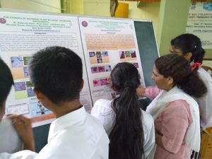
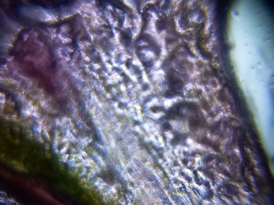
Infected leaf of a plant grown at the site of Nala passing through Kings Way Camp behind the Guru Teg Bahadur Khalasa College, University of Delhi
 Oct 06, 2018 • 12:43 AM UTC
 Unknown Location
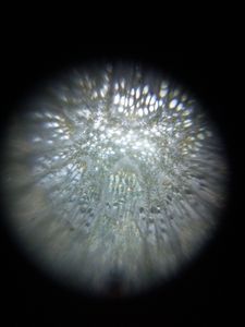
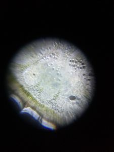
 140x Magnification
 Microorganisms
Kalawati saini
Learn about the author...
11posts
0comments
1locations
View in Media Gallery
T.S. of infected leaf. there are no clear cells boundaries (CELL WALLS). The stometa also was not seen. Our UV-VISIBLE ABSORPTION SPECTROSCOPIC data shows that the amount of leaf pigments( Chlorophyll, Carotenoid, Anthocynine) was very less then the same plant leaf grown in the garden of our college, DU This post is open to read and review on The Winnower.
Sign in to commentNobody has commented yet... Share your thoughts with the author and start the discussion!

 0 Applause
 0 Comments