Magic of stem cells!!
 Nov 18, 2014 • 1:39 AM UTC
 United States
 140x Magnification
 Unknown
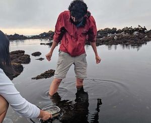
Manu Prakash
I am a faculty at Stanford and run the Prakash Lab at Department of Bioengineering at Stanford University. Foldscope community is at the heart of our Frugal Science movement - and I can not tell you how proud I am of this community and grassroots movement. Find our work here: http://prakashlab.stanford.edu
266posts
1198comments
42locations
View in Media Gallery
Summary: A simple prep to start looking at the wonders of developmental and stem cell biology – Live imaging of fly ovaries using Foldscope.
We owe a lot of our knowledge in biology to tiny little flies. As a model system goes; they have been remarkable in terms of genetic tools we have available now. So you might be wondering, if flies are so well understood – is it a crank that you churn and you get answers.
In short, No. But what makes science exciting is you have to find a way to map a “question” you care about to a “system” that you can ask the question, without loosing the jist of the question. Let’s say – you want to learn about stem cells. All multi-cellular animals have some kind of cells that remain undifferentiated. So in flies as well, these cells are found everywhere and give rise to many new structures. Here I will describe a simple technique, Bill Sullivan from UC Santa Cruz taught me – how to watch fly eggs develop and see germ line stem cells.
Methods :
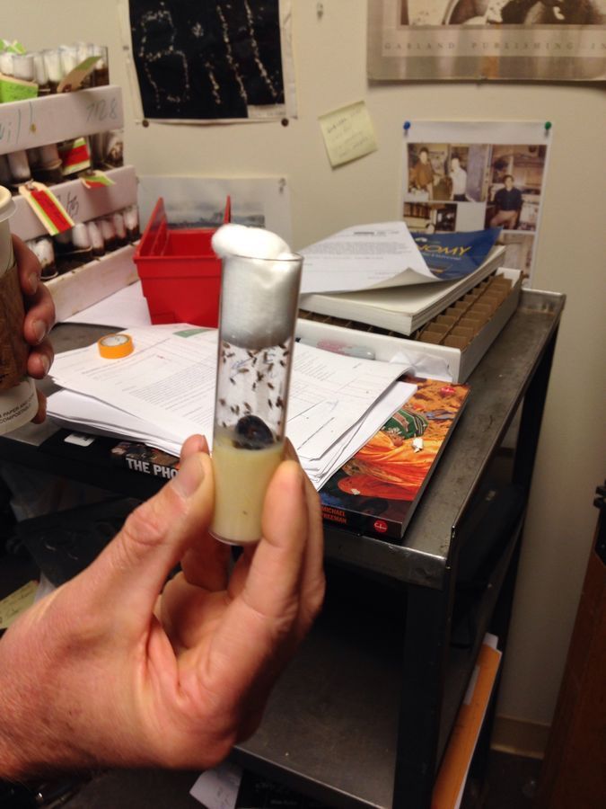
1. First you need to catch some flies. Any flies would do. Bill happened to have a fly stock, but I will start fresh here – so I caught some flies today morning. I posted a picture below (need to identify the same).
2. Now if you have ever tried to catch a fly, you know how fast they are. So you have to calm them down – literally. A simple trick I use is to cool them – yes put the ziplock bag in the freezer for 30seconds, or just out a ice cube on the fly. They will stop moving. I use this trick for all my insect surgeries.
3. Now it’s time to take the ovaries and developing eggs out. Take a shallow dish and fill it with water – so you can keep the organs you get out of the fly carefully, and not let them dry. Take two tweezers (any tweezers would do) and hold the abdomen and the back end of the fly with the two tweezers and just pull.. Splat.. All the innards including the gut will spill out. Ignore everything else for right now (or save it for later experiments – lots of surprises hiding in a fly).
** biology trick – biologists are proud of keeping good care of some simple tools. This includes a pair of tweezers, a nice hair-line brush and some safety pins. I also add some ziplock bags and a flash light to this list – who knows what you will find walking around.
4. You should see an organ that looks like a bunch of grapes. That’s what you really want – that’s where the germ line stem cells are hiding. Separate this organ out (fairly visible) and spread it on your tape/plastic slide.
5. Now we are ready to image. I will use both my low mag (140X Foldscope) and high mag (500X mag Foldscope) – since I know lots of things to see both at low and high mag.
6. The type of slide we just made – with a little bit of water is called a “wet mount”. This allows the biological samples to stay wet and thus not change morphology too much. Since we are going to image immediately, this will also be live imaging. Live imaging truly brings out colors of life, so you are up for a treat.
7. On purpose, I decided not to add any chemicals to color the sample. Although the sample is very transparent (and does not absorb much light); it actually provides for a fantastic contrast because of subtle mis-alignment of a few microns between the light source and the objective. This we get a phase-like image with fantastic contrast where I did not expect any contrast.
We owe a lot of our knowledge in biology to tiny little flies. As a model system goes; they have been remarkable in terms of genetic tools we have available now. So you might be wondering, if flies are so well understood – is it a crank that you churn and you get answers.
In short, No. But what makes science exciting is you have to find a way to map a “question” you care about to a “system” that you can ask the question, without loosing the jist of the question. Let’s say – you want to learn about stem cells. All multi-cellular animals have some kind of cells that remain undifferentiated. So in flies as well, these cells are found everywhere and give rise to many new structures. Here I will describe a simple technique, Bill Sullivan from UC Santa Cruz taught me – how to watch fly eggs develop and see germ line stem cells.
Methods :
1. First you need to catch some flies. Any flies would do. Bill happened to have a fly stock, but I will start fresh here – so I caught some flies today morning. I posted a picture below (need to identify the same).
2. Now if you have ever tried to catch a fly, you know how fast they are. So you have to calm them down – literally. A simple trick I use is to cool them – yes put the ziplock bag in the freezer for 30seconds, or just out a ice cube on the fly. They will stop moving. I use this trick for all my insect surgeries.
3. Now it’s time to take the ovaries and developing eggs out. Take a shallow dish and fill it with water – so you can keep the organs you get out of the fly carefully, and not let them dry. Take two tweezers (any tweezers would do) and hold the abdomen and the back end of the fly with the two tweezers and just pull.. Splat.. All the innards including the gut will spill out. Ignore everything else for right now (or save it for later experiments – lots of surprises hiding in a fly).
** biology trick – biologists are proud of keeping good care of some simple tools. This includes a pair of tweezers, a nice hair-line brush and some safety pins. I also add some ziplock bags and a flash light to this list – who knows what you will find walking around.
4. You should see an organ that looks like a bunch of grapes. That’s what you really want – that’s where the germ line stem cells are hiding. Separate this organ out (fairly visible) and spread it on your tape/plastic slide.
5. Now we are ready to image. I will use both my low mag (140X Foldscope) and high mag (500X mag Foldscope) – since I know lots of things to see both at low and high mag.
6. The type of slide we just made – with a little bit of water is called a “wet mount”. This allows the biological samples to stay wet and thus not change morphology too much. Since we are going to image immediately, this will also be live imaging. Live imaging truly brings out colors of life, so you are up for a treat.
7. On purpose, I decided not to add any chemicals to color the sample. Although the sample is very transparent (and does not absorb much light); it actually provides for a fantastic contrast because of subtle mis-alignment of a few microns between the light source and the objective. This we get a phase-like image with fantastic contrast where I did not expect any contrast.
View in Media Gallery
Here are the flies I picked from the stock. This is Drosophila – a workhorse for biologist.
View in Media Gallery
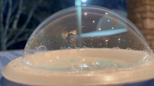
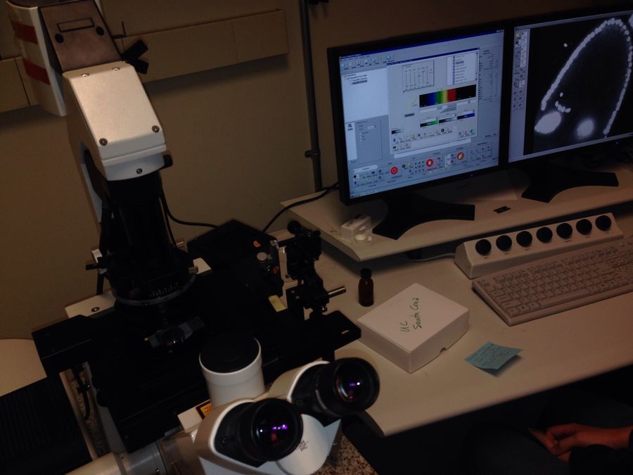
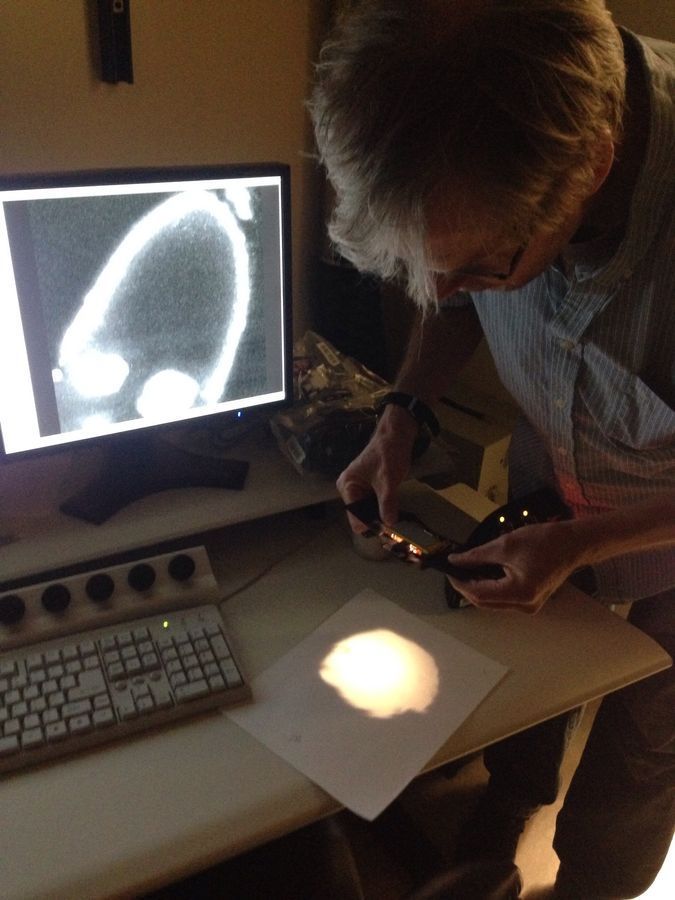
This is the usual confocal microscope bill uses, I put it for fun to show – what the commonly used tools look like.
View in Media Gallery
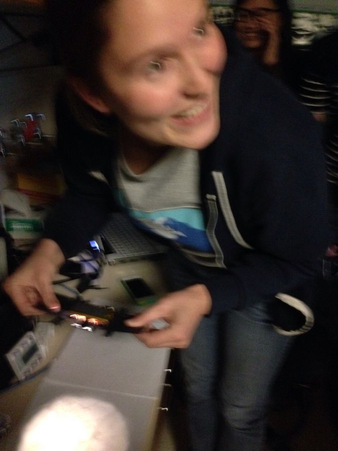
This was the moment the lab members in Bill’s fabulous lab realized that they could image all kinds of details – this was a really fun moment .
View in Media Gallery
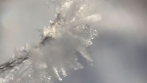
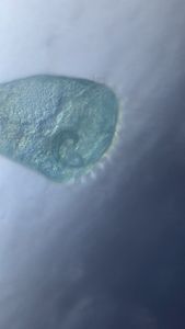
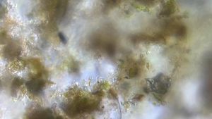
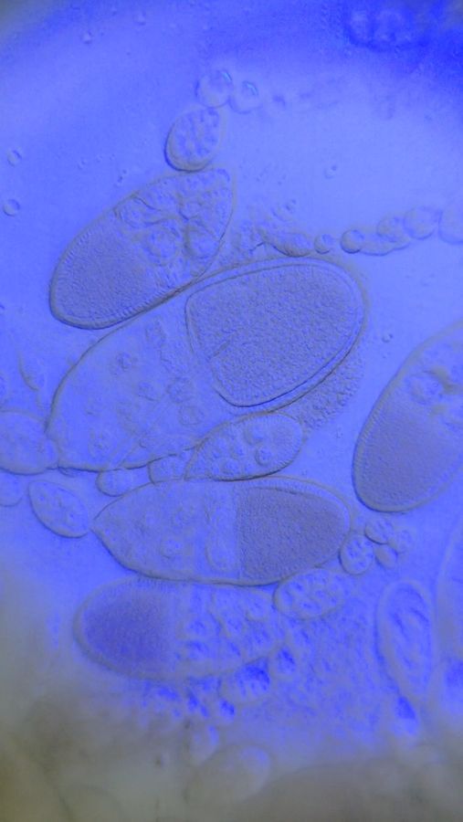
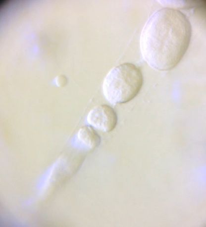
Low mag (140X) Foldscope, iphone 5
View in Media Gallery
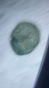
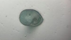
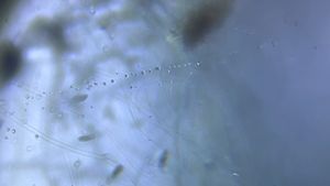
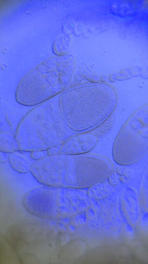
High mag (500X) Foldscope, iphone 5
Conclusions and next steps:
1. I am just so pleased with the images I was able to capture. I was easily able to identify Nurse cells, follicle cells and the cytoplasm of the main chamber of the egg.
2. The first thing to notice is that fly ovaries are like factories, they produce a series of eggs like a pipe line; and at the end of that pipeline is stem cells. Watch carefully at the end of this series of sequentially smaller egg chambers, and you will see a bundle of cells – those are germ line stem cells.
3. The follicle cells are similar to epithelial cells, covering the outside f the egg chamber.
4. I was expecting to see cytoplasmic streaming – so I want to mount more number of live flies to see f I can identify the same.
View in Media Gallery
Sign in to commentNobody has commented yet... Share your thoughts with the author and start the discussion!

 0 Applause
 0 Comments