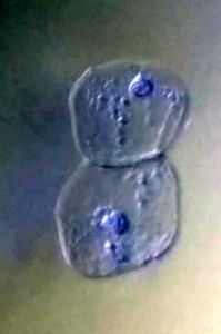
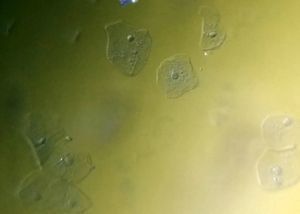
Cheek cells
 Jan 06, 2015 • 4:34 PM UTC
 Unknown Location
 140x Magnification
 Unknown
Laks Iyer
Human observer of life. https://sukshmadarshin.wordpress.com
97posts
1255comments
5locations
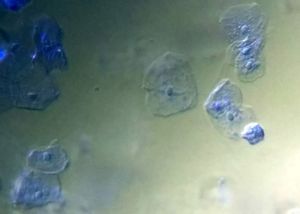
These are among the easiest samples to study. For this slide, I took a Q-tip/cotton bud and removed the cotton tip and swabbed my cheeks about 3-4 times gently. On a slide I put a drop of 1% Methylene Blue that I bought off the internet and mixed the end that I swabbed my cheek with in it a few times and added another drop of tap water. I then placed a cover slip and taped the edges.
I readily saw a large number of cheek cells floating around and the contrast between the nucleus and cytoplasm got better over time (3-5 minutes). Within the cytoplasm you see some granular material, perhaps rough endoplasmic reticulum. I also saw some bacteria floating around that are not visible in these frames. The condenser/LED system is really extraordinary, and I am sure with time I can get professional quality pictures (Work in progress). Need to try other stains.
Manu, what would you recommend for cleaning the lens.
Sign in to commentNobody has commented yet... Share your thoughts with the author and start the discussion!

 0 Applause
 0 Comments
_300x300.jpeg)


