Exploring the microscopic world of Bougainvillea -7 B
 Nov 01, 2018 • 9:10 AM UTC
 Unknown Location
 140x Magnification
 Microorganisms
kavitha sebastin
Learn about the author...
41posts
5comments
1locations
View in Media Gallery
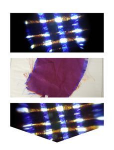
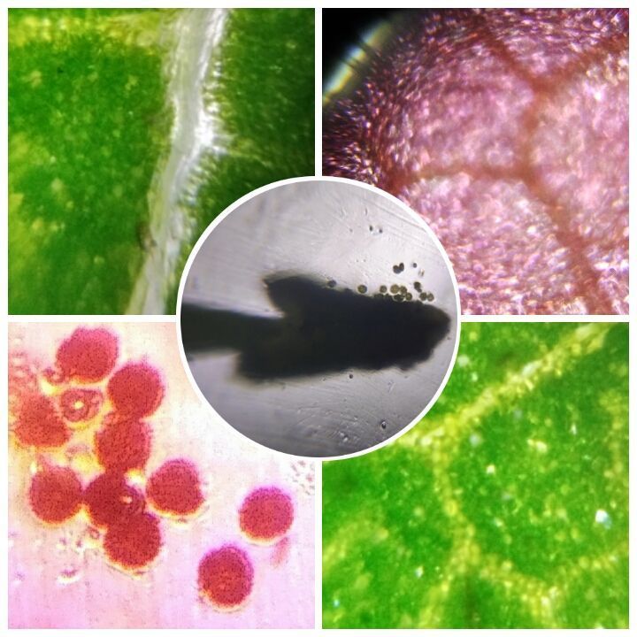
Veins,cells of coloured bract,anther,pollen grains and reticulate venation
View in Media Gallery
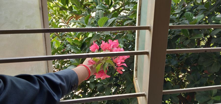
Bougainvillea twig with inflorescence near class window
View in Media Gallery
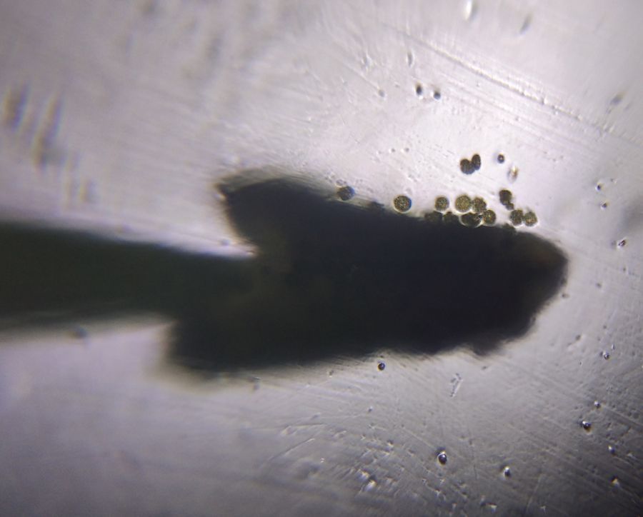
Anther releasing pollen grains
View in Media Gallery
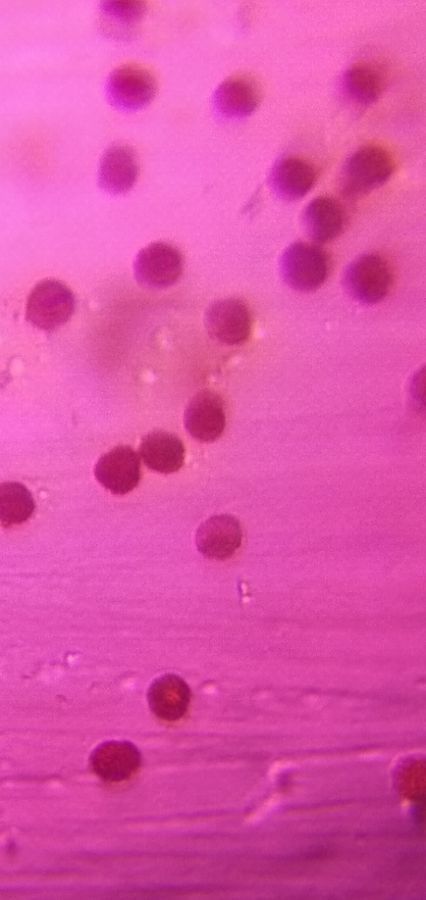
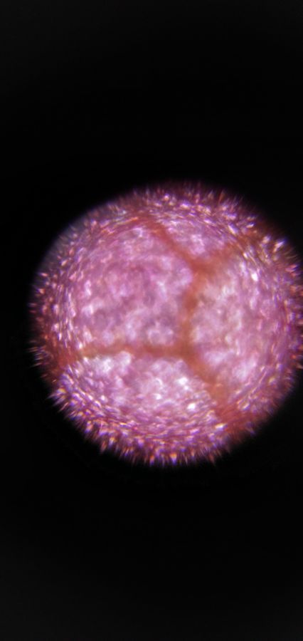
Pollen grains of Bougainvillea
View in Media Gallery
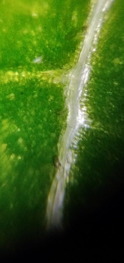
Veins of Bougainvillea leaf
View in Media Gallery
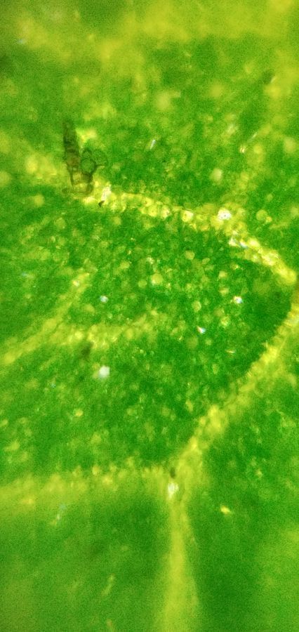
Leaf showing reticulate venation
View in Media Gallery
cells of bract of Bougainvillea Students of 7th were excited to see a branch of Bougainvillea plant bearing the inflorescence which had reached the second floor of their class window. The plant was creeping on a jack fruit tree.So taught of exploring the microscopic world of Bougainvillea flower and leaf through the foldscope.
Students observed the structure of anther releasing pollen grains,pollen grains, leaf cells showing veins and reticulate venation and the cells of the coloured bract using many foldscopes.
Students observed the structure of anther releasing pollen grains,pollen grains, leaf cells showing veins and reticulate venation and the cells of the coloured bract using many foldscopes.
Sign in to commentNobody has commented yet... Share your thoughts with the author and start the discussion!

 0 Applause
 0 Comments