Leaf surfaces of monocots
 Nov 06, 2018 • 4:37 AM UTC
 Unknown Location
 140x Magnification
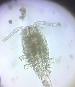
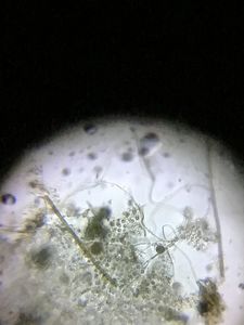
 Microorganisms
Jayashree Ramadas
We are a group of students, volunteers and staff working with TIFR Hyderabad's Science Education and Outreach program: http://www.tifrh.res.in/~outreach/
39posts
26comments
2locations
View in Media Gallery
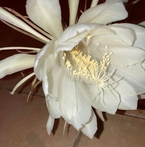
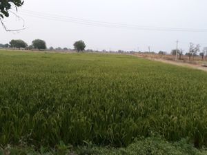
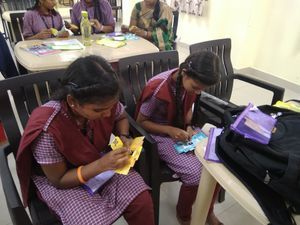
My sister’s home near Hyderabad has a beautiful garden with a number of ornamental and vegetable plants. While visiting her over a few days I decided to investigate the leaf stomata of monocot and dicot plants. In this post I will tell you about the monocots.
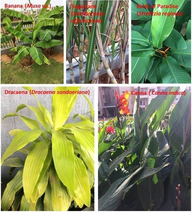
Among monocot plants I found Banana (Musa), Sugarcane (Saccharum), Birds of paradise (Strelitzia), Dracaena and Canna. We have been told that monocots have stomata on both the upper and the lower epidermis while dicots have them usually on the lower epidermis only — I decided to check this out.
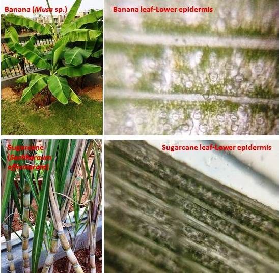
It turned out the lower epidermis in all these plant leaves (monocots and dicots) was easier to pull out with forceps than was the upper epidermis. A standard procedure is to use clear nail varnish to make an impression of the epidermis but, since I had no nail varnish, it took me a lot of effort. After many trials I was able to peel off the upper epidermis of Strelitzia, Dracaena and Canna, but the venation pattern of banana and sugarcane made the upper epidermis tear too easily, so I did not succeed till the end in peeling these off .
Among monocot plants I found Banana (Musa), Sugarcane (Saccharum), Birds of paradise (Strelitzia), Dracaena and Canna. We have been told that monocots have stomata on both the upper and the lower epidermis while dicots have them usually on the lower epidermis only — I decided to check this out.
It turned out the lower epidermis in all these plant leaves (monocots and dicots) was easier to pull out with forceps than was the upper epidermis. A standard procedure is to use clear nail varnish to make an impression of the epidermis but, since I had no nail varnish, it took me a lot of effort. After many trials I was able to peel off the upper epidermis of Strelitzia, Dracaena and Canna, but the venation pattern of banana and sugarcane made the upper epidermis tear too easily, so I did not succeed till the end in peeling these off .
View in Media Gallery
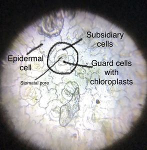
Monocot plants – Banana, Sugarcane, Strelitzia, Dracaena and Canna In Strelitzia, Dracaena and Canna I found that the lower epidermis had many more stomata than the upper epidermis. In the Banana, Sugarcane, Strelitzia and Canna lower epidermis I saw lots of stomata, arranged in regular linear patterns. The linear pattern in Dracaena was not so clear.

View in Media Gallery
Upper and lower epidermis of Birds of Paradise, Dracaena and Canna
View in Media Gallery
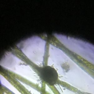
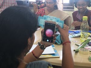
Lower epidermis of banana and sugarcane leaves Getting clear images of the epidermal surfaces was a challenge, and it took me many many trials to get all these photos. It helped that my first image, of the lower epidermis of Strelitzia, was beautifully clear. I immediately shared it on our TSWREIS Foldscope WhatsApp group and got some nice comments. This encouraged me to take up the extended project of exploring stomata.
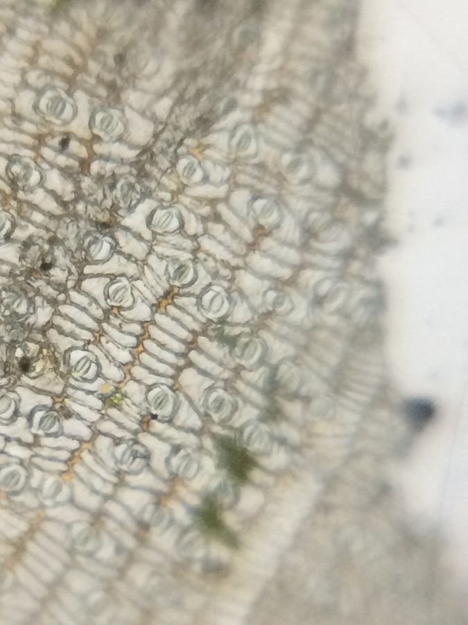
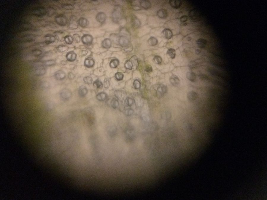
I did find the pattern that I expected, of stomata present both in upper and lower epidermis of monocots (for dicots, wait for the next post )! BUT the ‘Birds of paradise’ (Strelitzia) was an exception. In the image below see the lower epidermis which is fully crowded with stomata (my very beautiful slide!) but the upper epidermis shows very few or no stomata. I made about ten slides of the upper epidermis of different leaves of the same Strelitzia plant and of different plants too. Some of the cells were very small in size and perhaps the stomata were not clear. But it did look like stomata of Strelitzia are not equally distributed in the lower and upper epidermis and maybe this is an exceptional case among monocots.
I did find the pattern that I expected, of stomata present both in upper and lower epidermis of monocots (for dicots, wait for the next post )! BUT the ‘Birds of paradise’ (Strelitzia) was an exception. In the image below see the lower epidermis which is fully crowded with stomata (my very beautiful slide!) but the upper epidermis shows very few or no stomata. I made about ten slides of the upper epidermis of different leaves of the same Strelitzia plant and of different plants too. Some of the cells were very small in size and perhaps the stomata were not clear. But it did look like stomata of Strelitzia are not equally distributed in the lower and upper epidermis and maybe this is an exceptional case among monocots.
View in Media Gallery
Lower epidermis (showing a number of stomata) of Birds of Paradise (Strelitzia) leaves
View in Media Gallery
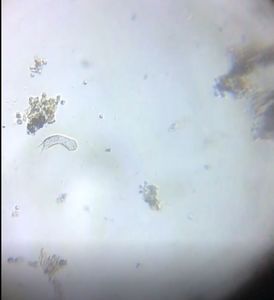
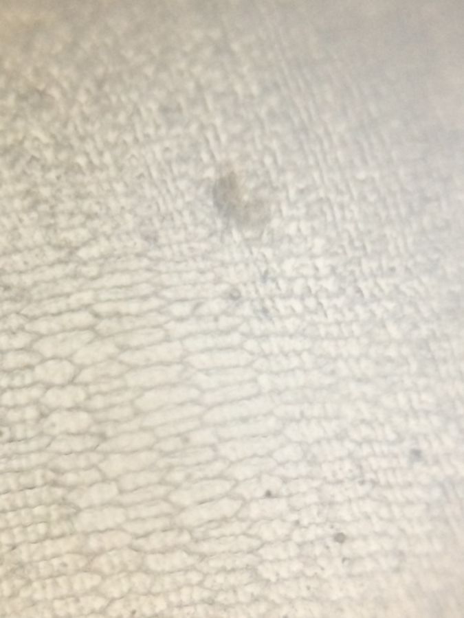
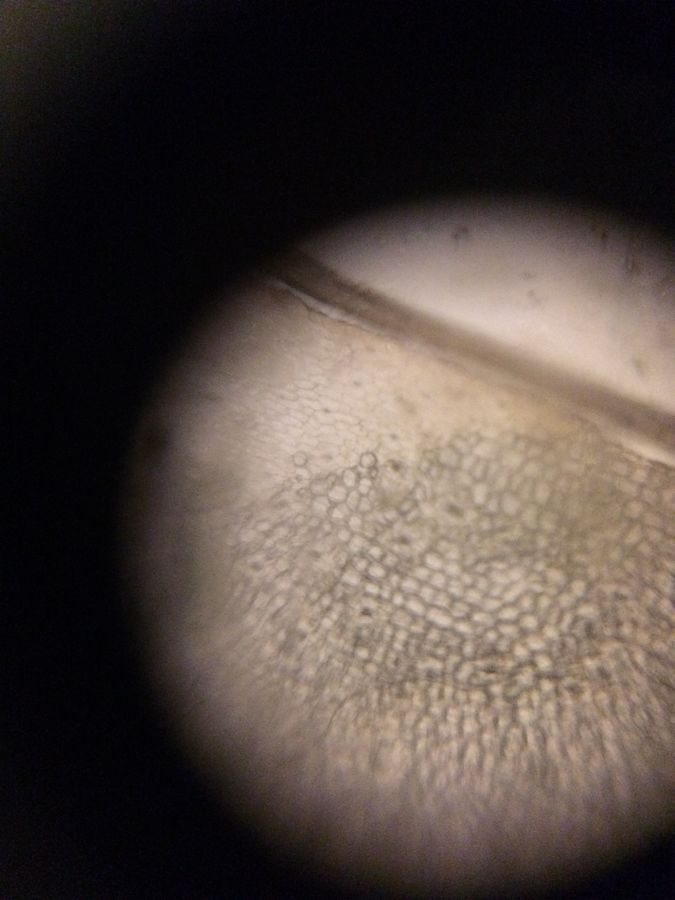
Upper epidermis of Birds of Paradise leaves Another surprise was that the guard cells in monocots are known to be in dumbbell shape but, in my specimen slides the shapes look bean shaped, not so different from dicots. Could it be a problem with my focusing? Please give your suggestions / comments!
View in Media Gallery
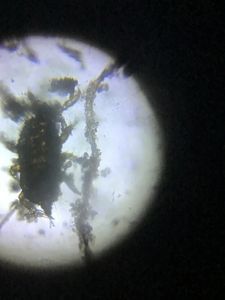
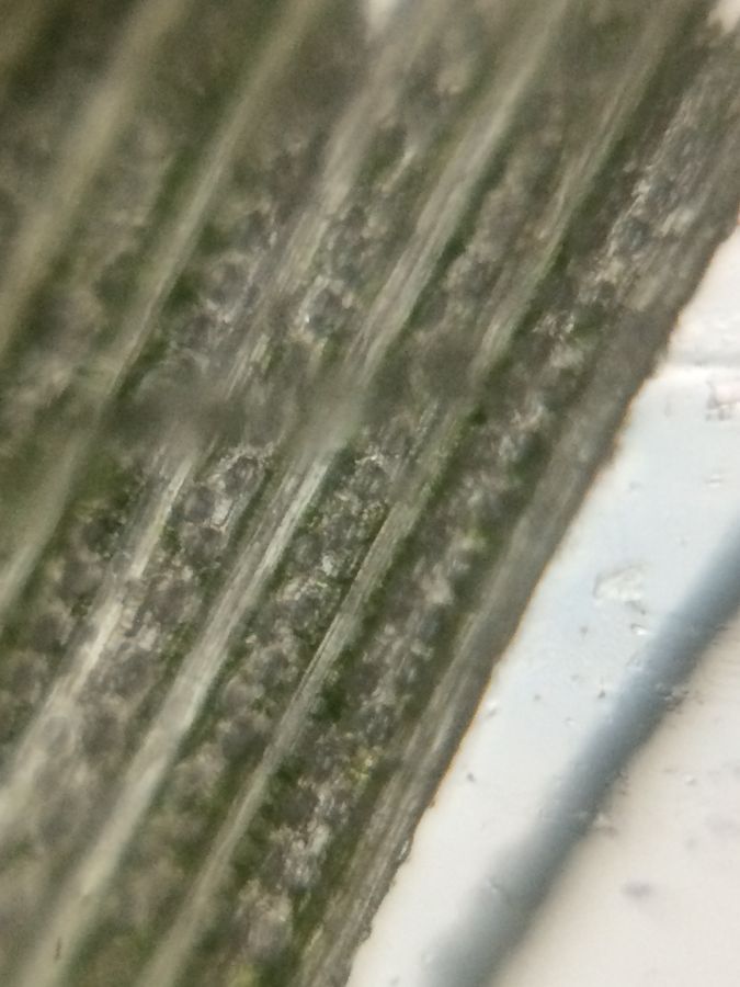
Sugarcane lower epidermis
View in Media Gallery
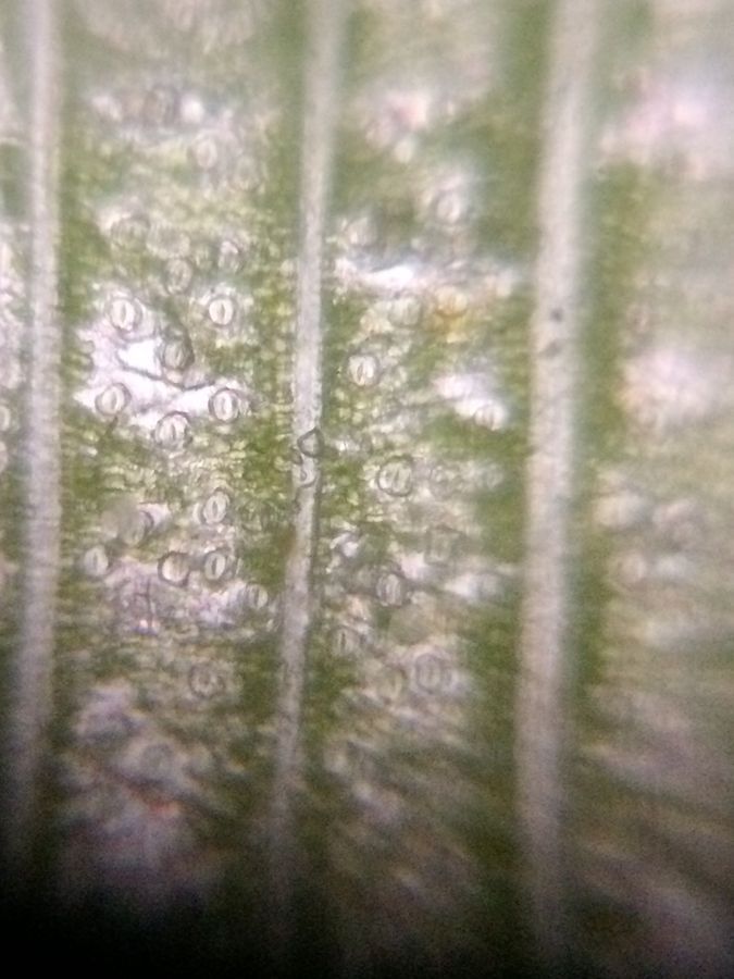
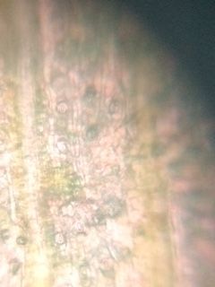
Banana lower epidermis
View in Media Gallery
Canna lower epidermis
View in Media Gallery
Dracaena lower epidermis
All my images are taken with an iPhone 5S A1530 with sunlight as the source. In one image of a banana leaf lower epidermis I saw a nice pink stain on the cells. Was it natural? I wondered. No, it was the colour of a couch which was in the direction of my viewing. A pretty slide it was.
All my images are taken with an iPhone 5S A1530 with sunlight as the source. In one image of a banana leaf lower epidermis I saw a nice pink stain on the cells. Was it natural? I wondered. No, it was the colour of a couch which was in the direction of my viewing. A pretty slide it was.
View in Media Gallery
Banana lower epidermis with pink light reflection I enjoyed making these slides and put in a lot of effort to get clear images of those slides. Now my photo app is fully loaded with just these images and my work with the epidermis of dicots (coming up in my next post!). To avoid confusion, I started with the photo of the plant, followed by upper and then lower epidermis. It took a number of deletions and additions of images to do a reasonable job.
Hope you all enjoyed this exploration of monocot leaf surfaces. In my next two posts, I will share my experience with some of the dicot leaf surfaces and a comparative view of both monocots and dicots . Till then, enjoy!!!
Ashalatha
(with Debashree and Jayashree)
Hope you all enjoyed this exploration of monocot leaf surfaces. In my next two posts, I will share my experience with some of the dicot leaf surfaces and a comparative view of both monocots and dicots . Till then, enjoy!!!
Ashalatha
(with Debashree and Jayashree)
Sign in to commentNobody has commented yet... Share your thoughts with the author and start the discussion!

 0 Applause
 0 Comments