Comparative view of monocot and dicot leaf surfaces
 Nov 06, 2018 • 4:37 AM UTC
 Unknown Location
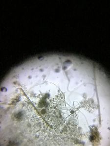
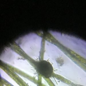
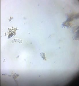
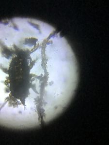
 140x Magnification
 Microorganisms
Jayashree Ramadas
We are a group of students, volunteers and staff working with TIFR Hyderabad's Science Education and Outreach program: http://www.tifrh.res.in/~outreach/
39posts
26comments
2locations

View in Media Gallery
View in Media Gallery
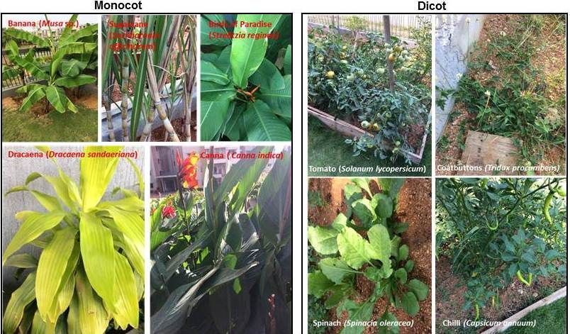
Monocot and dicot plants Working with the foldscope on nine different plants for about three days was a wonderful experience for me.
So, the final interpretations based on my observations are as follows:
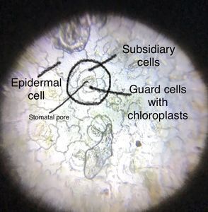
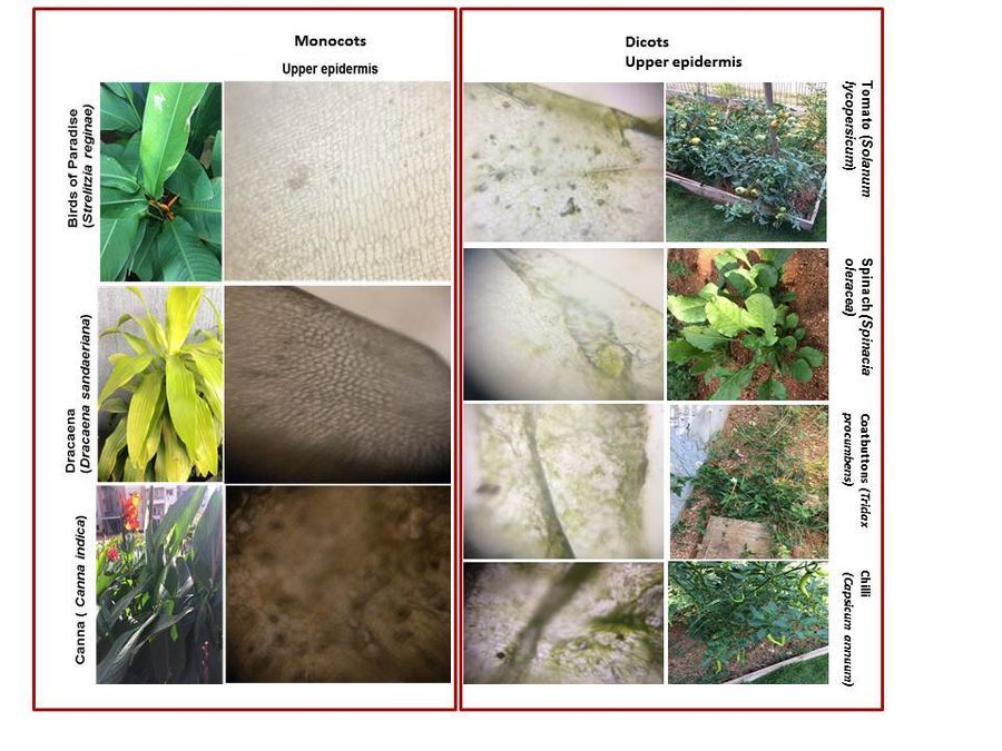
Monocot stomata are regularly arranged (e.g. Banana and Sugarcane), while dicot stomata are irregulalry arranged (e.g. Spinach, Tomato). Birds of Paradise plant may be an exception among monocots as the stomata are not equally distributed among the lower and the upper epidermis. Like monocots, in dicots too the upper epidermis possesses stomata. Here trichomes are produced in both lower and upper epidermis to avoid transpirational water loss through the stomata. I did not observe any trichomes in spinach (a dicot) nor in any of the monocots (but I wondered what made the surface of sugarcane leaves so rough). Here are some composite images showing the upper and lower epidermis of the monocots and dicots that I observed.
So, the final interpretations based on my observations are as follows:
Monocot stomata are regularly arranged (e.g. Banana and Sugarcane), while dicot stomata are irregulalry arranged (e.g. Spinach, Tomato). Birds of Paradise plant may be an exception among monocots as the stomata are not equally distributed among the lower and the upper epidermis. Like monocots, in dicots too the upper epidermis possesses stomata. Here trichomes are produced in both lower and upper epidermis to avoid transpirational water loss through the stomata. I did not observe any trichomes in spinach (a dicot) nor in any of the monocots (but I wondered what made the surface of sugarcane leaves so rough). Here are some composite images showing the upper and lower epidermis of the monocots and dicots that I observed.
View in Media Gallery
Upper epidermis of monocot and dicot plants

View in Media Gallery
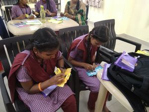
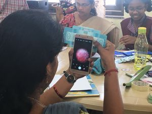
Lower epidermis of monocots and dicots While working on this short project I played with the foldscope for hours together to improve my technique while taking images. I still have to work a lot on that. I also followed the procedure from YouTube videos to take images with the phone but I had trouble with the coupler as it kept slipping out, which continuously distracted my concentration. Therefore, for all the images I placed the camera directly on the foldscope, focussed the field and then took the shot. It was laborious to handle, but my curiosity and interest with the foldscope encouraged me to continue my observations.
I would like to add a general observation that, the magnet of the coupler is too strong compared to the adhesive which attaches it to the camera. So the adhesive side of the coupler keeps slipping out. (I wonder if the adhesive softens in hot weather so people in tropical countries may notice it.) This makes it especially difficult while taking videos, when too I usually place the foldscope on the LED (without coupler) and keep the phone camera directly over it. This method works for me for taking photos as well. Please suggest if there is any alternative method to capture images without using the coupler!
Thanks you all.
Cheers!
Ashalatha
(with Debashree and Jayashree)
Sign in to commentNobody has commented yet... Share your thoughts with the author and start the discussion!

 0 Applause
 0 Comments