Watching spider eggs develop (14hr time lapse video)
 Aug 05, 2015 • 10:31 PM UTC
 Unknown Location
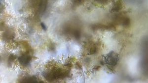
 140x Magnification
 Microorganisms
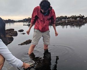
Manu Prakash
I am a faculty at Stanford and run the Prakash Lab at Department of Bioengineering at Stanford University. Foldscope community is at the heart of our Frugal Science movement - and I can not tell you how proud I am of this community and grassroots movement. Find our work here: http://prakashlab.stanford.edu
266posts
1198comments
42locations
View in Media Gallery
Babies are beautiful. Even for an organism like spider (which invoke mixed reaction in everyone); watching a tiny baby spider develop is incredibly beautiful. I get goose bumps talking about it. Also; I have been on a mission to image cell division using almost nothing but a foldscope (no specialized tools); and that led me to think about spiders.
Most recently – I did a small calculation about 20 million spiders in a small field. So where do all these spiders come from – eggs off ourse.
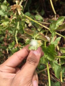
Spider eggs are fascinating. Firstly – they are everywhere (so they are trivial to run into in any garden). Secondly – they are mostly transparent enough to just watch them. So get out and get some spider egg sacs – put them in a foldscope and you get to watch them turning into little spider babies.
Methods:
0. First we have to collect some spider eggs. For a more detailed field survey and guide to collect spider eggs; see my prior post on collecting spider egg sacs in the field.
Here: http://microcosmos.foldscope.com/2015/07/26/20-million-spiders-lake-lagunita/
Most recently – I did a small calculation about 20 million spiders in a small field. So where do all these spiders come from – eggs off ourse.
Spider eggs are fascinating. Firstly – they are everywhere (so they are trivial to run into in any garden). Secondly – they are mostly transparent enough to just watch them. So get out and get some spider egg sacs – put them in a foldscope and you get to watch them turning into little spider babies.
Methods:
0. First we have to collect some spider eggs. For a more detailed field survey and guide to collect spider eggs; see my prior post on collecting spider egg sacs in the field.
Here: http://microcosmos.foldscope.com/2015/07/26/20-million-spiders-lake-lagunita/

1. I have been able to setup my foldscope on an old iPhone5 imaging using time lapse imaging. I was able to do 15 to 20 hour long continuous imaging that allows me to explore development of eggs.
2. I like using “lapse it” iPhone app since it gives me more control; buy you can also use the native iPhone time lapse.
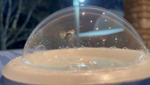
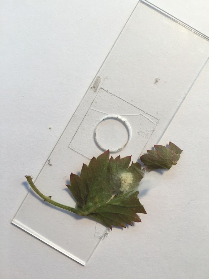
3. For the video below; I actually used foldscope in inverted mode. I remove the light module and just use the ambient light (using a table lamp in this case below). Inverted light imaging has the advantage of ease of setting up the organisms.
2. I like using “lapse it” iPhone app since it gives me more control; buy you can also use the native iPhone time lapse.
3. For the video below; I actually used foldscope in inverted mode. I remove the light module and just use the ambient light (using a table lamp in this case below). Inverted light imaging has the advantage of ease of setting up the organisms.
View in Media Gallery
4. To avoid evaporation and loss of water; I also put some wet cotton around the microscope.
5. As you can see in the time lapse below; the sunrise is clearly visible (with the advent of too much ambient light). Next time; I will close the curtains.
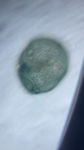
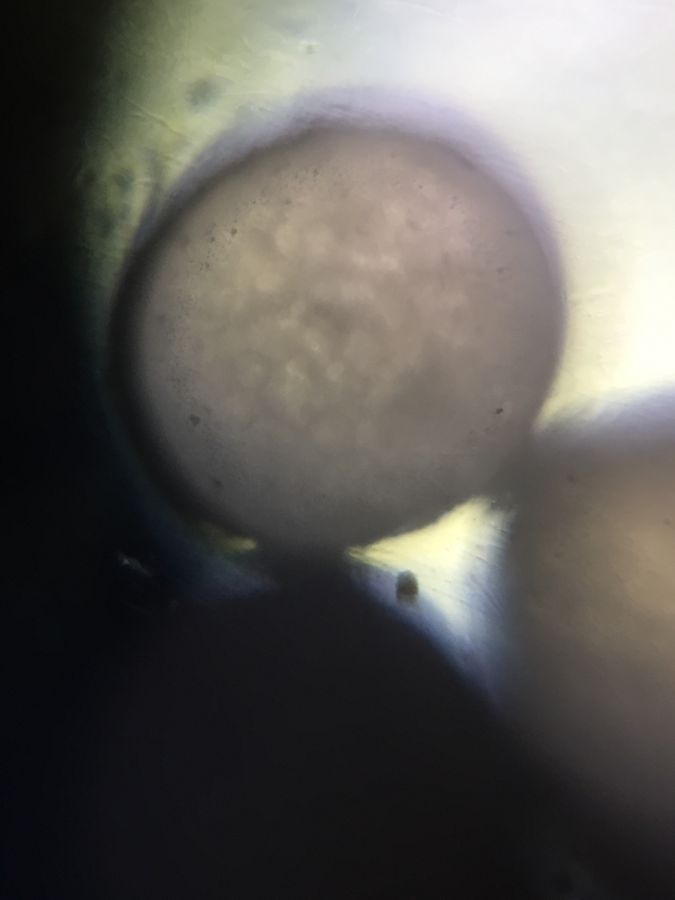
6. The eggs are almost pearl like – incredibly beautiful to look at. Also; in a single egg sac – I can get sychronized eggs which allows me to image them simultaneously.
5. As you can see in the time lapse below; the sunrise is clearly visible (with the advent of too much ambient light). Next time; I will close the curtains.
6. The eggs are almost pearl like – incredibly beautiful to look at. Also; in a single egg sac – I can get sychronized eggs which allows me to image them simultaneously.
Observations:
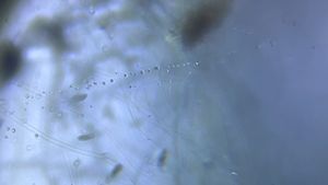
– the video above was collected for 14 hrs straight with 848 frames collected. I play them here at 40fps.
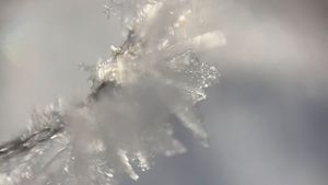
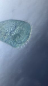
– it’s apparent that the egg is “alive” as you see cellular movements inside the egg. If you really look hard; you can see individual cell division events on the surface.
– a lot of yolk granules (round cell shaped objects) are clearly visible.
– the video above was collected for 14 hrs straight with 848 frames collected. I play them here at 40fps.
– it’s apparent that the egg is “alive” as you see cellular movements inside the egg. If you really look hard; you can see individual cell division events on the surface.
– a lot of yolk granules (round cell shaped objects) are clearly visible.
View in Media Gallery
– I can not say for sure if the egg was viable for the entire time of imaging. I need to improve my moist cotton ball technique to make sure no moisture is lost during imaging. So I will be trying these imaging setups with really long term imaging (36 or 48hrs straight). I am excited about that but also hoping nobody calls me during that time 🙂 ha ha.
The first time I ran this long term experiment; I did not realize the evaporation rate due to the eggs being exposed.
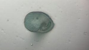
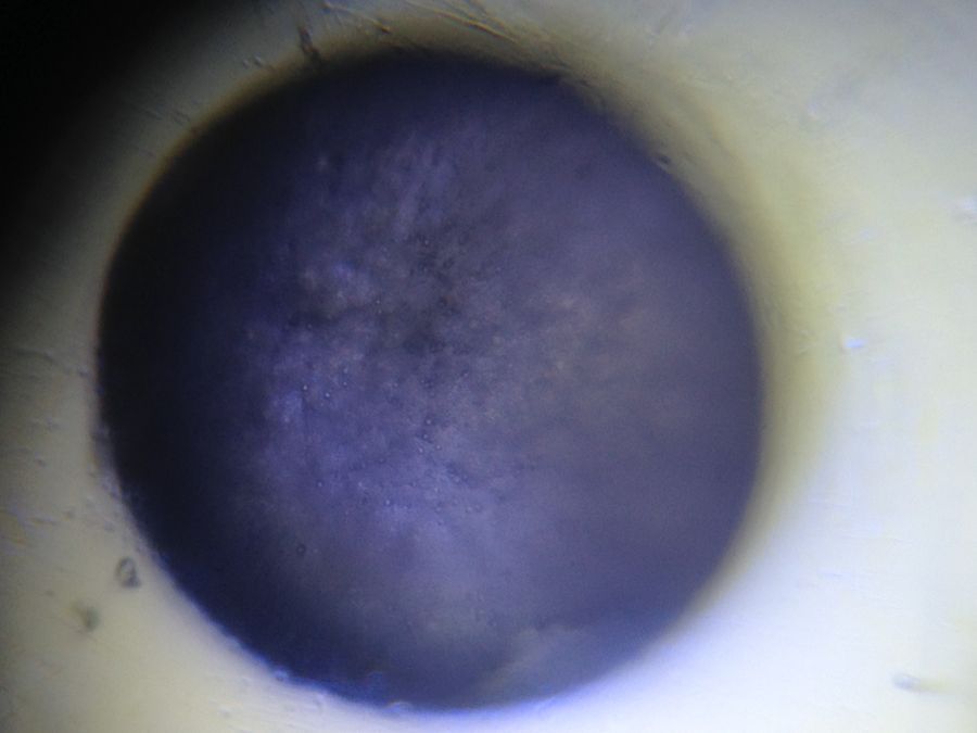
Here is another video of egg development in dark field setup. To configure your foldscope in dark field – please see a prior post by Matt..
http://microcosmos.foldscope.com/2015/06/24/simple-dark-field-improve-your-imaging-contrast-with-another-classic-microscopy-technique/
As you can see in the video below – desiccation (loss of water) deflates the egg unfortunately – making it non viable.
The first time I ran this long term experiment; I did not realize the evaporation rate due to the eggs being exposed.
Here is another video of egg development in dark field setup. To configure your foldscope in dark field – please see a prior post by Matt..
http://microcosmos.foldscope.com/2015/06/24/simple-dark-field-improve-your-imaging-contrast-with-another-classic-microscopy-technique/
As you can see in the video below – desiccation (loss of water) deflates the egg unfortunately – making it non viable.
Finally; although this is an ongoing experiment – it has convinced me of the real value of long term time lapse imaging. I am incredibly excited about the potential of watching life forms take shape and form. Give it a try; the reward is literally experiencing one of the most beautiful phenomenon in life – how do life forms take shape and form from a simple single cell to trillions of cells working together. Although I can’t answer that question as yet; at least I can watch this most wonderful phenomena in my kitchen.
Keep exploring.
Manu
ps: Although I do not know what spider species I have the eggs from; I did make a curious observation in my home office a few days ago. I had caught some spider that escaped; and I woke up one morning to find a beautiful spider web right on my home lab chair. What a beautiful web.

View in Media Gallery
37.8636586 -122.2500124
Sign in to commentNobody has commented yet... Share your thoughts with the author and start the discussion!
 0 Applause
 0 Comments