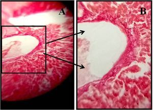
Photomicrographs of H & E stained Rat liver section –DEN induced hepatocarcinogenesis after Lithium Chloride treatment
 Nov 08, 2018 • 9:51 PM UTC
 Unknown Location
 140x Magnification
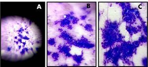
 Microorganisms
Ratna Kumari
Learn about the author...
39posts
3comments
1locations
View in Media Gallery
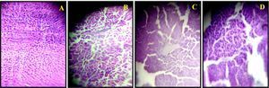
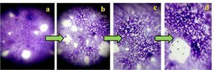
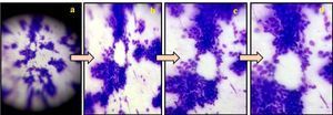
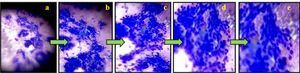
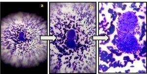
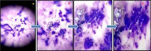
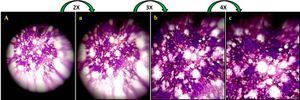
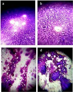
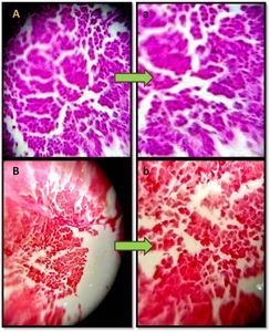
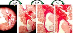
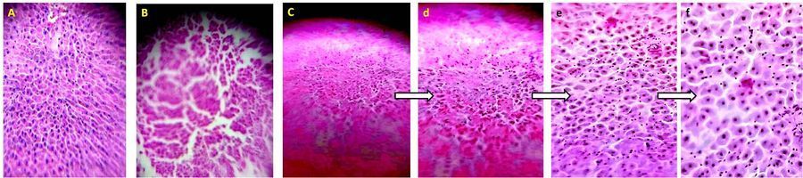
Photomicrographs of H & E stained Rat liver section –DEN induced hepatocarcinogenesis after Lithium Chloride treatment. Panel A shows untreated rat liver sections, with normal liver architecture, cuboidal hepatocytes, and radiating hepatic cords surrounding the central vein. Panel B shows DEN treated rat liver sections, with disorganized liver architecture, dilated sinusoids, and expanded portal vein. Panel C and d,e,f shows post Lithium Chloride treated rat liver sections with cuboidal hepatocytes like untreated rat liver sections. d, e, f are the zoom out images of image C, which were taken on OPPO-F7 android cell phone. d is 1.2X , e is 2.5X and f is 4X zoom out.
Sign in to commentNobody has commented yet... Share your thoughts with the author and start the discussion!
 0 Applause
 0 Comments