Mosquito larvae
 Sep 07, 2015 • 12:17 PM UTC
 Unknown Location
 140x Magnification
 Microorganisms
Saad Bhamla
Learn about the author...
32posts
11comments
2locations
View in Media Gallery
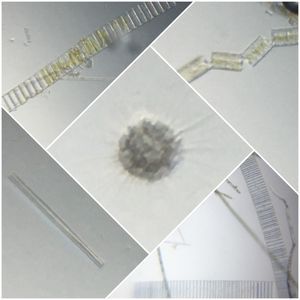
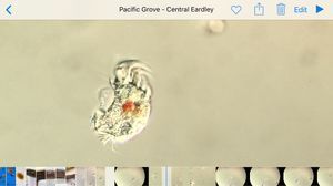
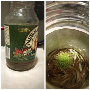
I came across some mosquito larvae in the lab and decided to image them using the foldscope (with tom’s help).
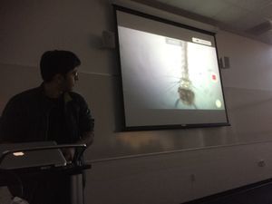
Some of the videos have a running commentary, which may add to your experience (in my opinion) as I discuss my observations with fellow lab-mates – it’s always nice to have a few extra pairs of eyes.
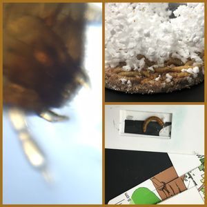
So. Here’s a simple video showing these creatures swimming around in a tub of tap-water. The wire mesh is used to scoop them out for preparing on a slide.
http://youtu.be/a9t_5aAc3ho
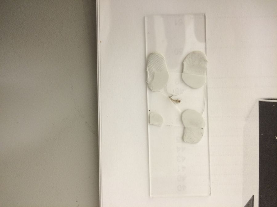
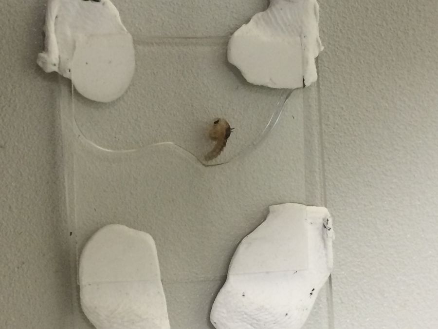
I deposited a live larvae onto a glass slide, added some spacers, a few drops of water, and gently pressed a glass coverslip on top of the specimen. Be careful not to press to hard as the coverslip breaks easily!!
Some of the videos have a running commentary, which may add to your experience (in my opinion) as I discuss my observations with fellow lab-mates – it’s always nice to have a few extra pairs of eyes.
So. Here’s a simple video showing these creatures swimming around in a tub of tap-water. The wire mesh is used to scoop them out for preparing on a slide.
http://youtu.be/a9t_5aAc3ho
I deposited a live larvae onto a glass slide, added some spacers, a few drops of water, and gently pressed a glass coverslip on top of the specimen. Be careful not to press to hard as the coverslip breaks easily!!
View in Media Gallery
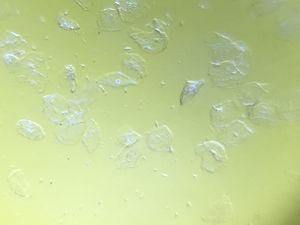
Now for the fun part. In the next three videos below, you’ll see the mosquito larvae images using the foldscope. We observed their breathing apparatus, fine hair near their mouth, gut movements, muscle twitching, and feeding motions.
Video 1:
http://youtu.be/KtEQ4eR8XZo
Video 2:
http://youtu.be/mnwHNH2fuMQ
Video 3:
http://youtu.be/IAnE3fAZ_ZY
Video 4:
http://youtu.be/yicyFWnQ-aM
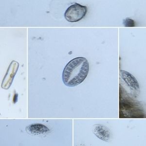
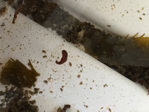
Next we prepared a mosquito pupae. In the image below, you should see a distinct shape difference compared to the previous image of the larvae stage.
Video 1:
http://youtu.be/KtEQ4eR8XZo
Video 2:
http://youtu.be/mnwHNH2fuMQ
Video 3:
http://youtu.be/IAnE3fAZ_ZY
Video 4:
http://youtu.be/yicyFWnQ-aM
Next we prepared a mosquito pupae. In the image below, you should see a distinct shape difference compared to the previous image of the larvae stage.
View in Media Gallery
And here’s a video of the pupae stage.
http://youtu.be/ZEY8WOWwwEI
Finally, I captured some slow motion videos of the larvae swimming around.
http://youtu.be/VZBRqQwm7sU
http://youtu.be/ZEY8WOWwwEI
Finally, I captured some slow motion videos of the larvae swimming around.
http://youtu.be/VZBRqQwm7sU
Sign in to commentNobody has commented yet... Share your thoughts with the author and start the discussion!

 0 Applause
 0 Comments