Holotrich Protozoa identification in a field setting
 Jun 06, 2019 • 1:59 AM UTC
 Unknown Location
 140x Magnification
 Microorganisms
Meignanalakshmi Sundaram
I am Dr.S.Meignanalakshmi, working as Professor, at the Directorate of Centre for Animal Health Studies, TANUVAS, Chennai-51. Working on Foldscope project on "Foldscope for diagnosis of Rumen Acidosis and parasitic infections in cattle" sanctioned by DBT
66posts
8comments
1locations

View in Media Gallery
DBT Foldscope scheme – “ Foldscope for diagnosis of Rumen acidosis and Parasitic infection in cattle”.
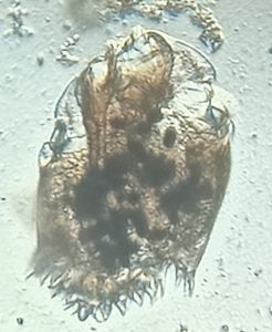
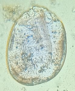
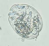
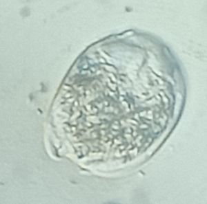
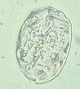
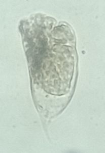
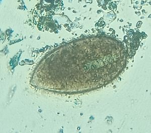
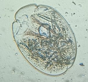
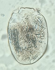
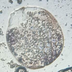
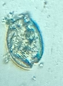
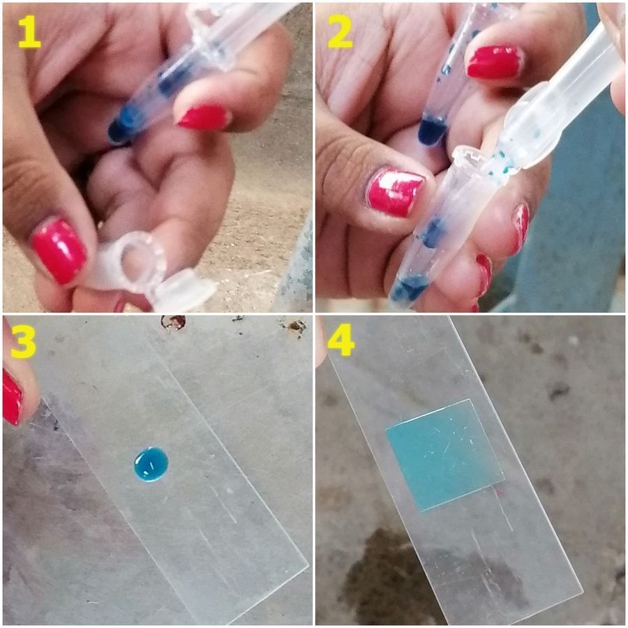
This post is in response to Manu Prakash’s comment on our previous post (Holotrich protozoa – from Rumen fluid). Rumen fluid was collected from an healthy cow (Figure 1) in a sterile container (figure 2). A drop of rumen fluid was taken and 5 to 10 times the volume of MFS (methyl green Formalin Saline) stain was added ( figure 3). After an incubation time of about 30 mins, the mixture was taken and viewed under Foldscope (Figure 4).
This post is in response to Manu Prakash’s comment on our previous post (Holotrich protozoa – from Rumen fluid). Rumen fluid was collected from an healthy cow (Figure 1) in a sterile container (figure 2). A drop of rumen fluid was taken and 5 to 10 times the volume of MFS (methyl green Formalin Saline) stain was added ( figure 3). After an incubation time of about 30 mins, the mixture was taken and viewed under Foldscope (Figure 4).
View in Media Gallery
Figure 1 – Collection of Rumen fluid

View in Media Gallery
Figure 2: Rumen fluid collected in a sterile container
View in Media Gallery
Figure 3: MFS staining in field setting

View in Media Gallery
Figure 4: Slide viewed under Foldscope
Sign in to commentNobody has commented yet... Share your thoughts with the author and start the discussion!

 0 Applause
 0 Comments