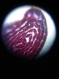
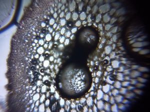
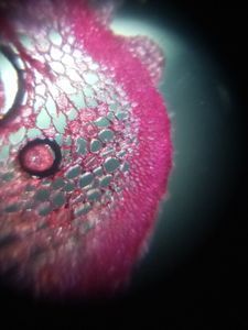
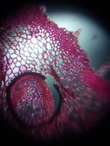
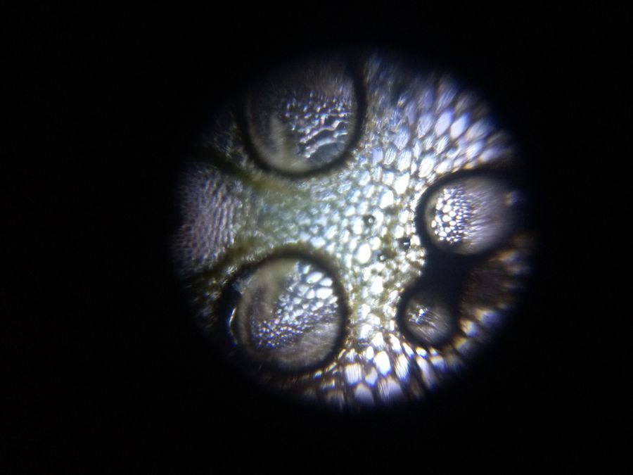
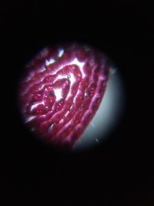
T. S. of Fern Petiole
 Nov 30, 2018 • 9:27 PM UTC
 Unknown Location
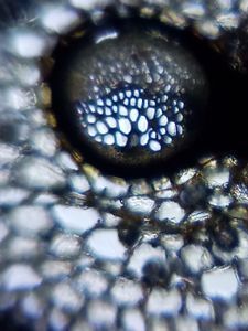
 140x Magnification
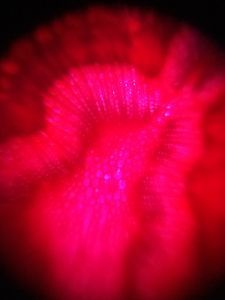
 Microorganisms
Kalawati saini
Learn about the author...
15posts
0comments
1locations
View in Media Gallery
T.S. of PETIOLE of Fern without staining., Phloem tissue is made sieve tubes, companion cells are phloem parenchyma.
Sign in to commentNobody has commented yet... Share your thoughts with the author and start the discussion!
 0 Applause
 0 Comments