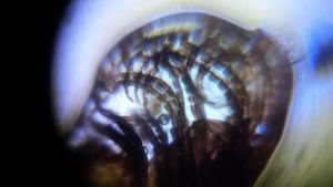
C. elegans in bright field and dark field
 Oct 07, 2015 • 7:42 AM UTC
 Unknown Location
 140x Magnification
 Microorganisms
Tom Hata
Learn about the author...
17posts
18comments
3locations
View in Media Gallery
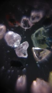
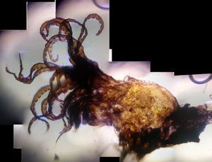
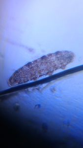
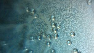
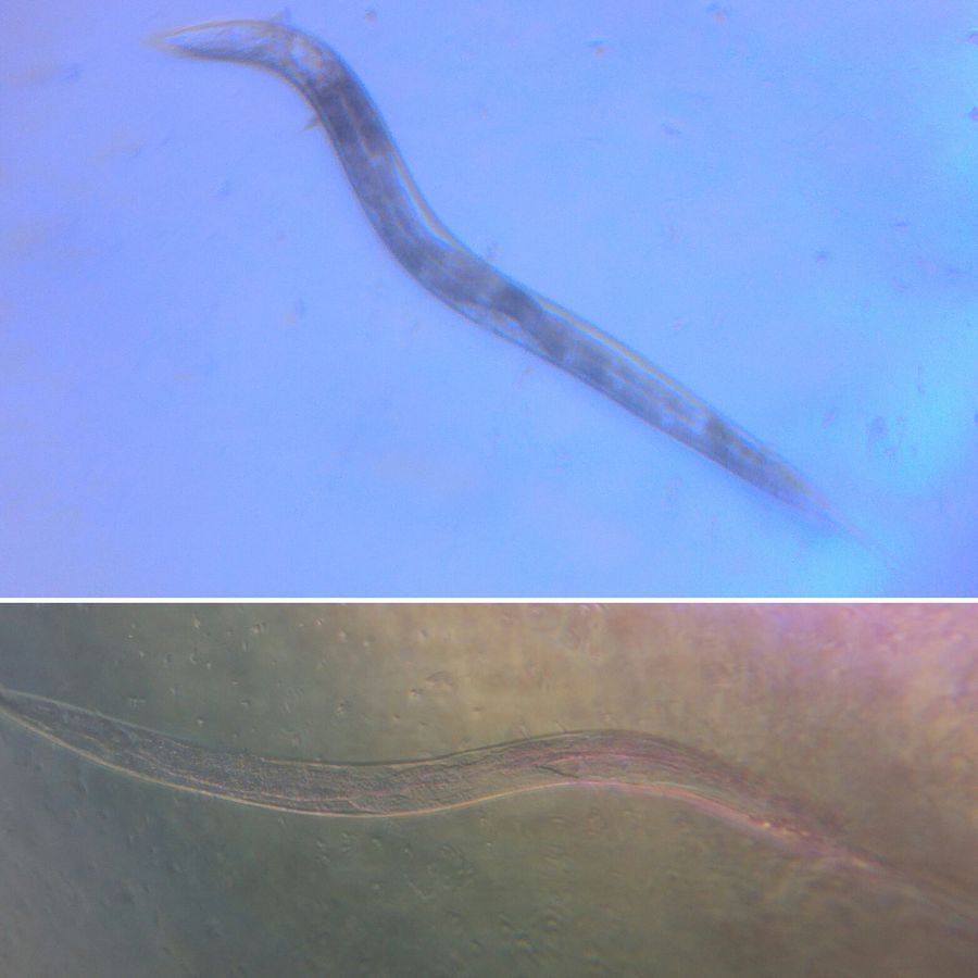
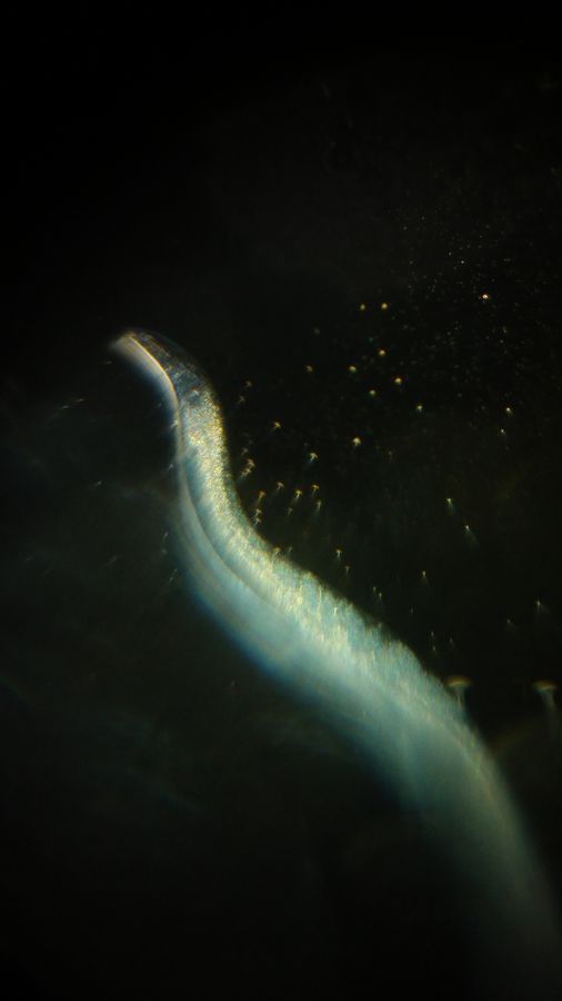
Thanks to an ongoing experiment, the lab is flooded with animals! The animal du jour is everyone’s favorite model organism, the nematode C. elegans. The trouble with imaging C. elegans under the Foldscope is that they are (1) very small, (2) mostly transparent, and (3) highly mobile (Most of the time. See the video at the end for an example of not-so-elegans swimming). This makes tracking an individual on a glass slide a daunting task. One trick to increasing the contrast of transparent organisms is using dark-field microscopy. I used a method described in a previous post (https://microcosmos.foldscope.com/?p=7042) to achieve this. As this is my first pass at this technique, this post serves as evidence of my trials and tribulations. The image above distinctly shows the difference between an image taken in bright field (top) and dark field (bottom). It was much easier to locate C. elegans on the slide in dark field, as its features exhibited much greater contrast. The image below is… somewhere in the middle of the previous two, although it was an attempt at dark field. I’m guessing I needed to make ink dot on the tape much darker to obtain a stereotypical dark-field image, although the specimen’s features do contrast nicely. Tomorrow will be another day to refine this technique… more to come.
View in Media Gallery
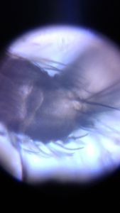
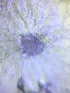
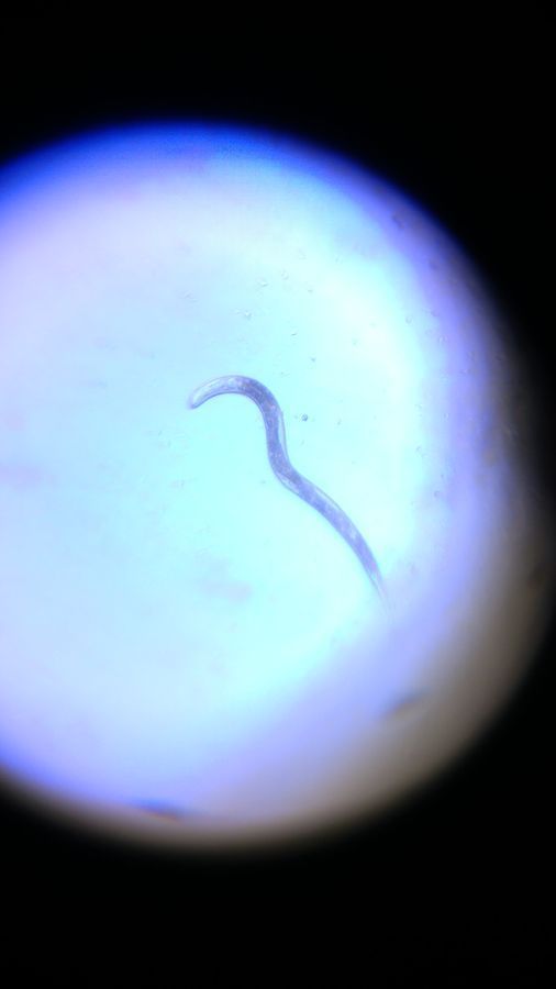
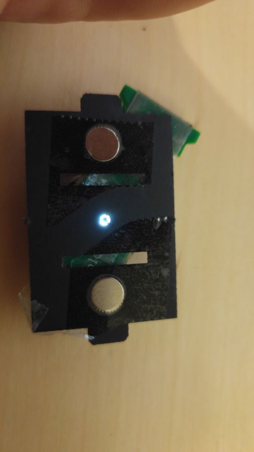
UPDATE: Turns out you can get much, much better dark field results with a paper aperture.
View in Media Gallery
I cut this piece on the laser cutter, leaving the middle piece attached by 3 thin strips. I usually shy away from superlatives, but the resultant images absolutely blew my mind. Even after dozens upon dozens of Foldscoping, I haven’t even begun to scratch the surface of this tool’s potential! For videos of dark field in action, see below.
View in Media Gallery
Sign in to commentNobody has commented yet... Share your thoughts with the author and start the discussion!
 0 Applause
 0 Comments