Importance of well adjusted illumination: using a flower to study a flower
 Oct 11, 2015 • 12:18 AM UTC
 Unknown Location
 140x Magnification
 Microorganisms
Manu Prakash
I am a faculty at Stanford and run the Prakash Lab at Department of Bioengineering at Stanford University. Foldscope community is at the heart of our Frugal Science movement - and I can not tell you how proud I am of this community and grassroots movement. Find our work here: http://prakashlab.stanford.edu
266posts
1198comments
42locations
View in Media Gallery
Note: this is a continuing post on a strange flower I had stumbled upon – if you have not seen the earlier post; try taking a look here first.
Light transmission through flower petals
Lookin at an organism with the help of the same organism.
This is going to be a cyclic post. We are going to use a biological organism to study itself. What I mean by that – is I will use a flower petal as a diffuser in the foldscope to image its own cells and figure out why it makes for such a great diffuser. I am already smiling – just thinking about it.
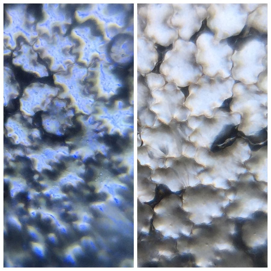
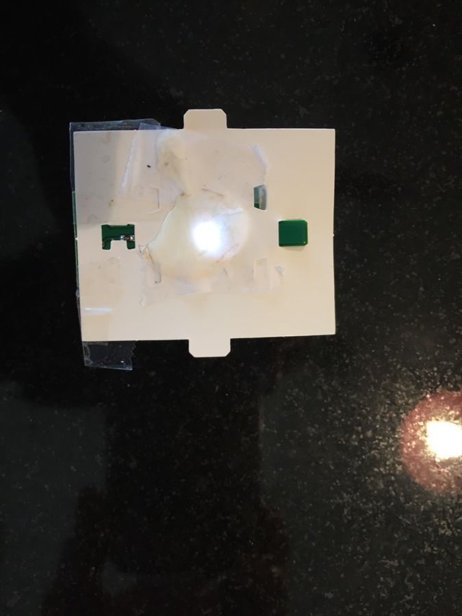
First; here is the demonstration to show why you need to think carefully and put together your illumination unit. It’s always valuable to compare – and that’s why Foldscope has a very open, modular structure. The image below is the same flower petal imaged with and without a diffuser between the LED light and the sample (on the bottom side of the Foldscope).
Light transmission through flower petals
Lookin at an organism with the help of the same organism.
This is going to be a cyclic post. We are going to use a biological organism to study itself. What I mean by that – is I will use a flower petal as a diffuser in the foldscope to image its own cells and figure out why it makes for such a great diffuser. I am already smiling – just thinking about it.
First; here is the demonstration to show why you need to think carefully and put together your illumination unit. It’s always valuable to compare – and that’s why Foldscope has a very open, modular structure. The image below is the same flower petal imaged with and without a diffuser between the LED light and the sample (on the bottom side of the Foldscope).

View in Media Gallery
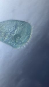
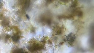
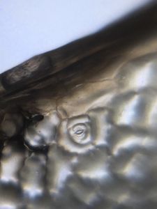
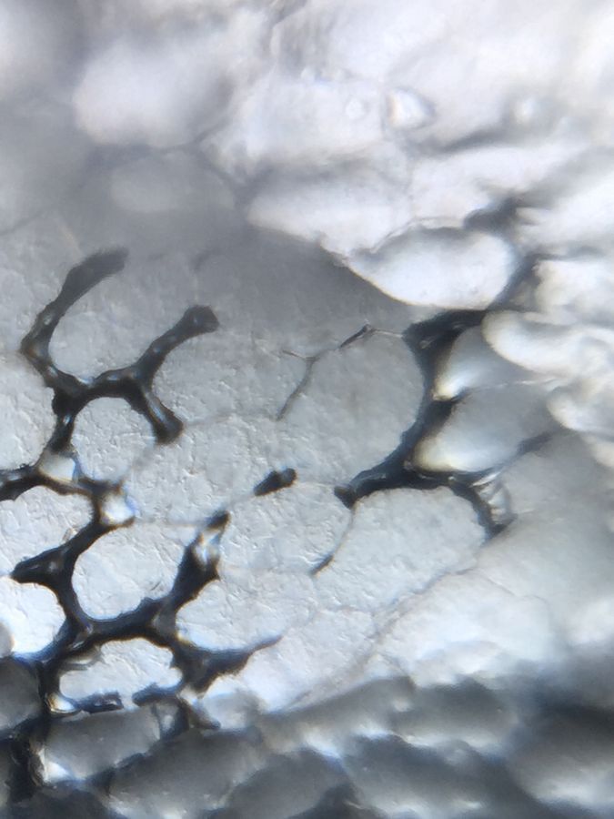
To the left is flower petal cells (buttercup) where I did not use any diffuser between the LED and the same. On the right – I played a clever trick – where I use tw flower petal itself as a diffuser. This is what the diffuser looks like.
View in Media Gallery
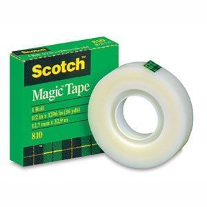
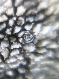
Now; you can use many different type of diffusers like a simple tissue paper or diffusion tape (by 3M). The “invisible magic tape” by 3M is a nice diffuser. But the flower petal is a even better diffuser. This reduces chromatic aberrations significantly and you see internal skeletal structures inside the cells; that are completely invisible in the first image.
View in Media Gallery
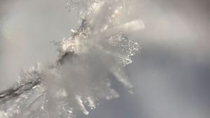
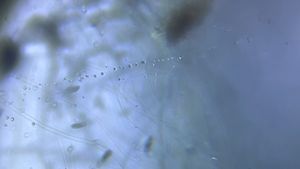
Here is a quick movie for you to enjoy beautiful cytoskeleton structures inside these curved cells.
Now; I was able to setup a time lapse on my foldscope and also watch these curved cells pop as a function of time. Here is a 8 sec movie where every frame was taken every 5 min and run for 2hrs.
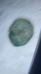
Now; I am also excited about finding stomata on flower petals. I did not think about this before; but all flower petals seem to have some degree of stomata. If you have some insight on function of stomata on flower petals – please do drop a note below.
Now; I would love for anyone to suggest a live stain that I could use to stain the skeletal like fibers I see inside the petal cells. They seem to keep the cells pegged up and curved.
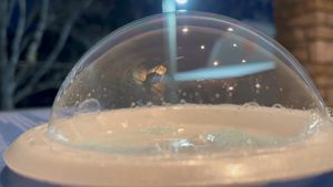
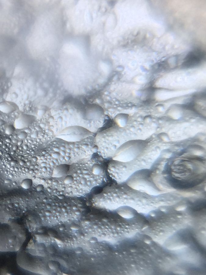
I have also noticed that over time; a lot of moisture actually collects on the petals themselves. They themselves act as little lenses.
I have also noticed that over time; a lot of moisture actually collects on the petals themselves. They themselves act as little lenses.
View in Media Gallery
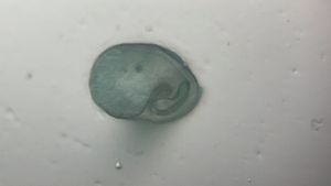
Also, I can squash the cells and flatten them up to get to see structures inside the cells as well. Take a look.
View in Media Gallery
Also; @Laks recent pointed about a paper that has looked at reflection properties of butter cups. Here is a link to the same.
“How does buttercup light up your chin”
http://www.ncbi.nlm.nih.gov/pmc/articles/PMC3350741/
So; next time you are Foldscoping – think about your illumination and never shy away from optimizing your illumination.
Keep exploring
Manu
“How does buttercup light up your chin”
http://www.ncbi.nlm.nih.gov/pmc/articles/PMC3350741/
So; next time you are Foldscoping – think about your illumination and never shy away from optimizing your illumination.
Keep exploring
Manu
Sign in to commentNobody has commented yet... Share your thoughts with the author and start the discussion!
 0 Applause
 0 Comments