Histology of regenerating earthworm
 Dec 09, 2018 • 8:46 PM UTC
 Unknown Location
 140x Magnification
 Microorganisms
Jackson Durairaj Selvan Christyraj
Learn about the author...
13posts
0comments
1locations
View in Media Gallery
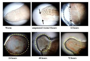
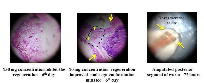
Figure 2: The above figures (a, b) show the histology of regenerating worm. a.) Inhibition of regeneration was observed at a high dose of triphala (150 mg/ml). b.) Triphala in 10mg/ml concentration are able to induce the regeneration capability of earthworm on 6 th of post-amputation. c.) The worm amputated at the 30 th segment is unable to regenerate their anterior portion. Arrow denotes the amputation sites (10 th & 30 th segment respectively). *denotes the formation of new segments.
View in Media Gallery
Sign in to commentNobody has commented yet... Share your thoughts with the author and start the discussion!

 0 Applause
 0 Comments