A food web in a fluid droplet (algae, rotifers, and anemone)
 Oct 21, 2015 • 9:39 PM UTC
 Unknown Location
 140x Magnification
 Microorganisms
Tom Hata
Learn about the author...
17posts
18comments
3locations

View in Media Gallery
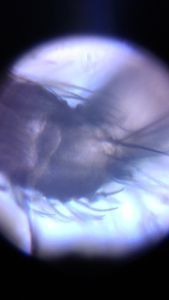
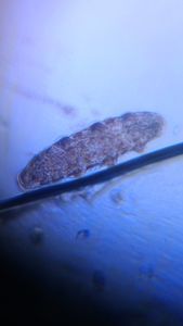
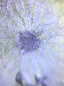
In my ongoing quest to explore marine microfauna, I recently observed rotifers under a Foldscope. Like many motile marine invertebrates, rotifers use cilia to maneuver through the water column. They also use these cilia to feed by generating currents.

View in Media Gallery
The anterior end of the animal is in the top right of the pictures, and there are 2 crowns of cilia on either side (this image in high mag). The cilia on these crowns beat in waves that generate current, allowing locomotion and feeding. Please excuse the psychedelic color changes in the first video, this was due to the light source oversaturating my camera phone’s sensor. I remedied this by using several layers of Scotch brand magic tape to diffuse the light.


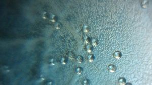
High magnification was necessary to even see the cilia beating, as they appeared as a blur in low magnification. For scale, those little spheres of algae are approximatey 4 microns in diameter. I was curious to see whether I could observe the ciliary waves by filming it in high speed. Here’s a video of the cilia beating, filmed at approximately 130 fps and slowed down 8x. Although I wasn’t able to get the cilia entirely in focus, there are a number of curious things happening. First, the algae provide an excellent set of particles to visualize the flow patterns created by the cilia. Second, the rotifer would periodically stop feeding and would retract. I’m wondering if this is because it has caught a particle, or if there is some other reason for this behavior.
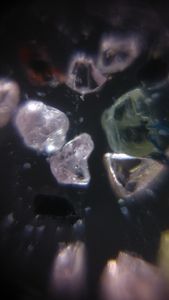
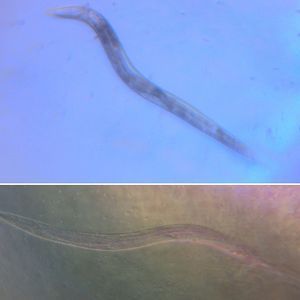
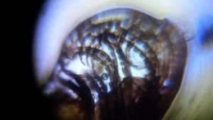
BONUS! I was also able to get my hands on a few anemone polyps (seen here in dark field and low mag). These polyps were so tiny that I was able to just barely fit them on a microscope slide, with 2 pieces of double-sided tape used as spacers.

View in Media Gallery
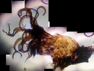
My challenge was to see whether I could record an anemone capturing and feeding on a rotifer. After a few hours and a dozen trials, success was at hand. Below is an anemone with a rotifer trapped in its mouth (the round shape on the right end), about to be consumed.
View in Media Gallery
In fact, I was able to capture the process of consumption over a 6-minute span.
By expanding and contracting, it looks like the anemone forces the rotifer down using a combination of cilia and peristaltic movement. At the end, you can even see the intact rotifer in the anemone’s gut! Here’s the same video, sped up 24x to get a better sense of the overall ingestion process.
Sign in to commentNobody has commented yet... Share your thoughts with the author and start the discussion!
 0 Applause
 0 Comments