Carpobrotus – a pig face plant
 Oct 27, 2015 • 4:22 AM UTC
 United States
 140x Magnification
 Microorganisms
Manu Prakash
I am a faculty at Stanford and run the Prakash Lab at Department of Bioengineering at Stanford University. Foldscope community is at the heart of our Frugal Science movement - and I can not tell you how proud I am of this community and grassroots movement. Find our work here: http://prakashlab.stanford.edu
266posts
1198comments
42locations
View in Media Gallery
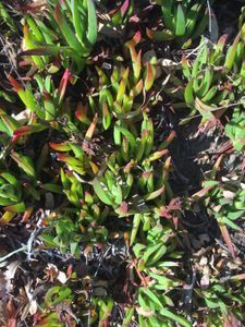
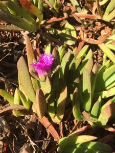
Its a little bit ironic for a plant to be called pig face. Specially when it looks like a beautiful flower. This is a very common plant – carpobrotus – with almost 25 or so species spread all across the coast of California. It would be rare to be at a beach in California and not run into this beautiful succulent. Although, it is a native of South Africa, it feels right at home in California or Australia.
But to survive in the harsh salty environment of the beach or California coast; you need some special features. Every time I saw this plant; I thought about what’s so special for it to proliferate like wild fire. So I decided to run a foldscope project to explore what turned to be an incredible beauty. Nothing about this plant is pig face; so here I submit my case to rename this plant – call it anything but not a pig face please.
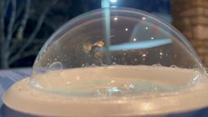
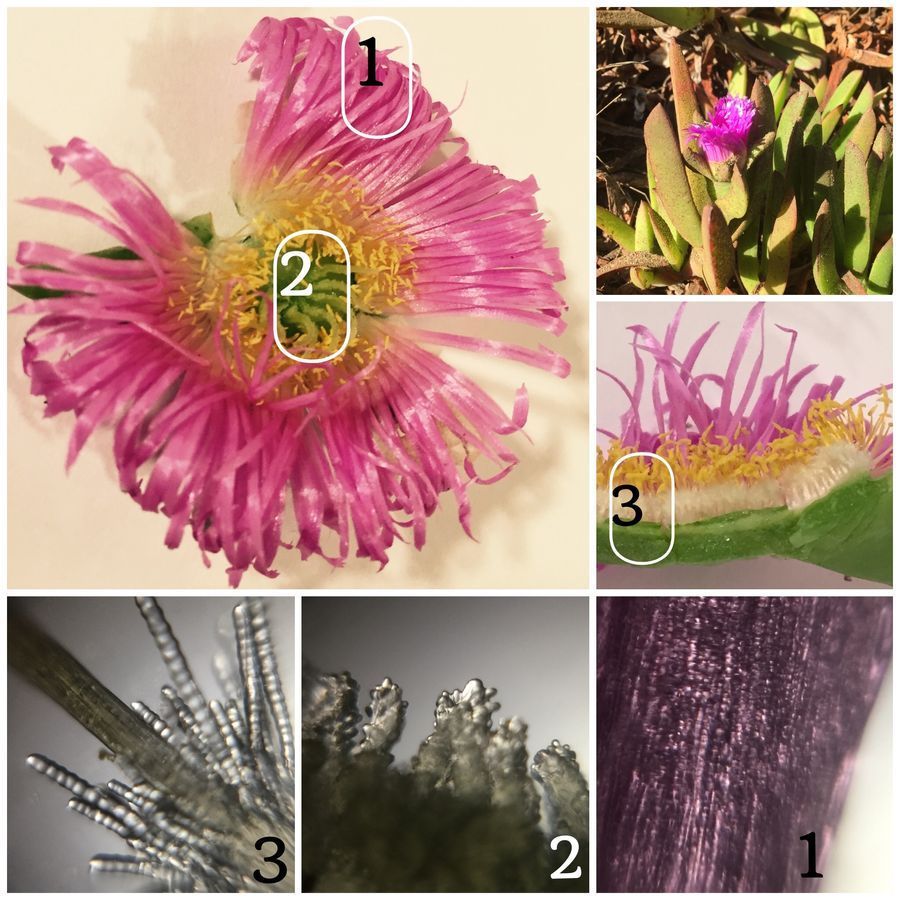
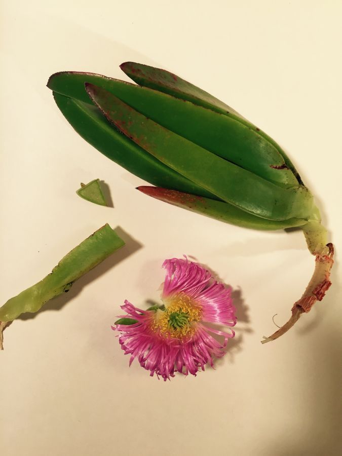
The first feature I noticed is that the leaves were succulent – means full of water. The moment you break a little bit of the leaf; you realize it is filled with water. I made some slices to take a look at the same. Also, I manage to find a beautiful pink flower which I will explore here in great detail.
But to survive in the harsh salty environment of the beach or California coast; you need some special features. Every time I saw this plant; I thought about what’s so special for it to proliferate like wild fire. So I decided to run a foldscope project to explore what turned to be an incredible beauty. Nothing about this plant is pig face; so here I submit my case to rename this plant – call it anything but not a pig face please.
The first feature I noticed is that the leaves were succulent – means full of water. The moment you break a little bit of the leaf; you realize it is filled with water. I made some slices to take a look at the same. Also, I manage to find a beautiful pink flower which I will explore here in great detail.
Here is what Indecuded to explore; a flower and its succulent leaf. Notice the amount of water in the leaf – it’s almost as if the plant knows what it means to live in a desert. With a California drought – it gives great meaning to savings water for the future. Who knows when is going to rain again in California (no pun intended).
View in Media Gallery
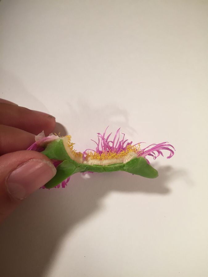
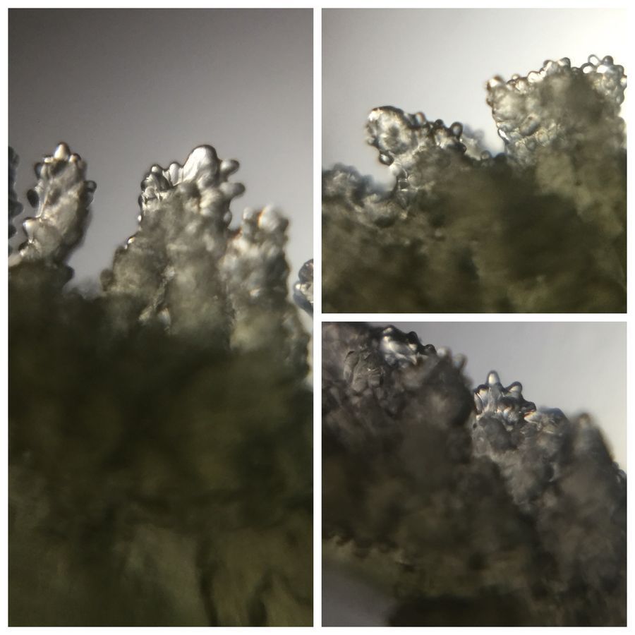
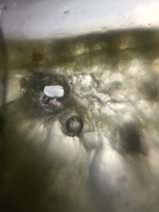
So, now it was time to look at the flower up close in a foldscope. The first I ipened up the flower and noticed a very strange white brush like growth at the bottom of the anther.
View in Media Gallery
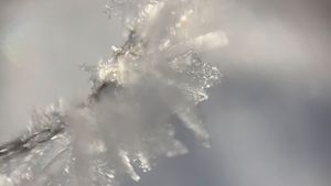
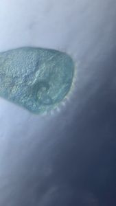
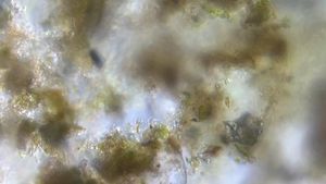
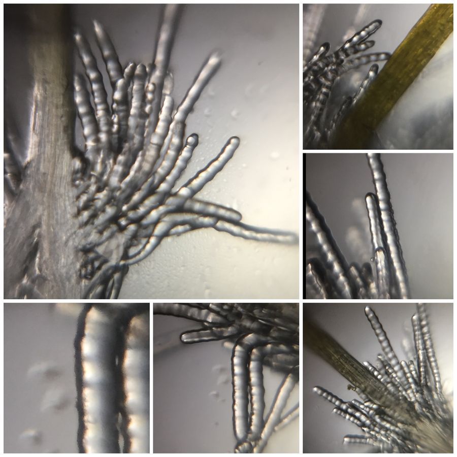
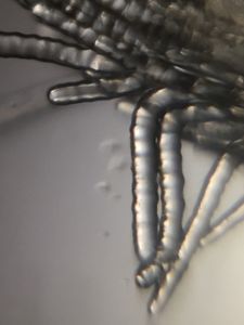
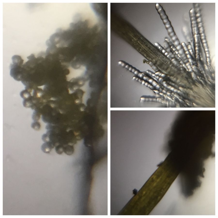
It turned out, the flower has some beautiful trichomes. I imaged them for a while; here is a short collection of the same.
View in Media Gallery
I was so excited about the trichomes; I was able to actually take a time lapse video of the Trichomes drying. Since I had broken the anther stalk from the flower – it dried under the microscope for 4 hours. By taking an image every 30 seconds; here I captured a really fun video of the Trichomes drying and collapsing on itself.
As can be seen, the Trichomes collapse on themselves as they dry. I Also noticed sub cellular structures inside the Trichomes – but inwas not able to capture them well with no stains.
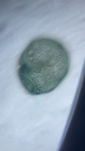
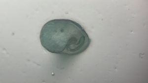
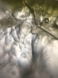
Next, I focused my attention on a green structure in the middle of the flower which seems to form a strane bulbulous curved structure. Here are few foldscope images of the same.
View in Media Gallery
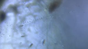
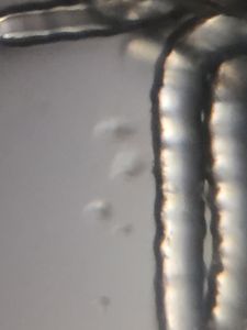
It appeared to me that this surface receives the pollen grains. So next I looked for the pollen grains and in a minute or so, I was indeed able to image the same.
View in Media Gallery
Finally, I also looked at the water filled leaves. They mostly were water – but to my surprise; I found as intricate a cellular structure as is found in common leaves. Some kind of vasculature and plumbing to move all that water around.
I can not think about this plants capacity to handle salt water. Now, if someone can comment and let me know that this plant is not toxic, I would like to see what the water in the leaves actually taste like. Until that time; I will eagerly wait for the much needed rains in California.
Cheers
Manu
Ps: if you have specific questions or would like me to image a specific portion of plant; please leave your requests below.
Cheers
Manu
Ps: if you have specific questions or would like me to image a specific portion of plant; please leave your requests below.
Sign in to commentNobody has commented yet... Share your thoughts with the author and start the discussion!
 0 Applause
 0 Comments