Onion Cell Stains
 Aug 13, 2022 • 1:46 PM UTC
 United States
 140x Magnification
 Plants
Holly Stuart
Learn about the author...
35posts
15comments
8locations
View in Media Gallery
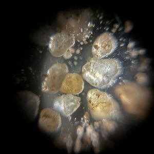
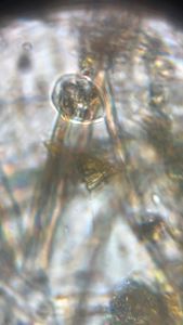
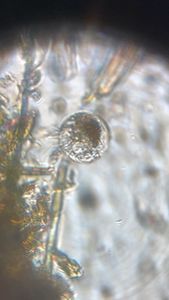
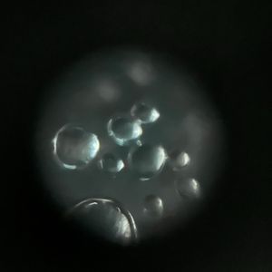
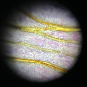
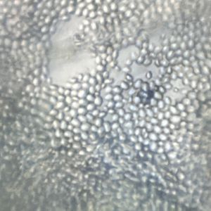
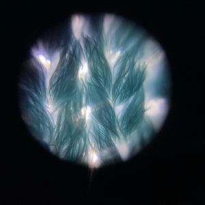
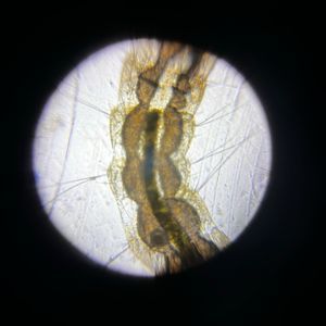
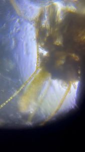
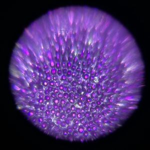
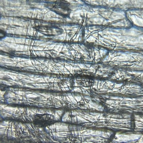
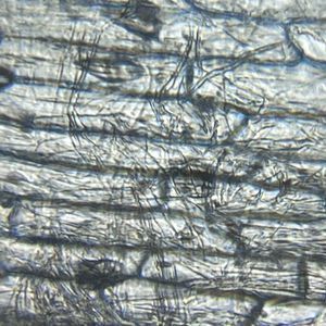
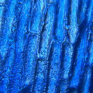
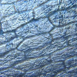
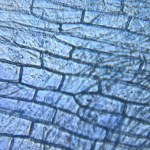
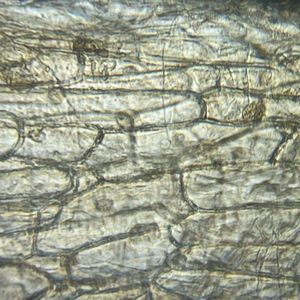
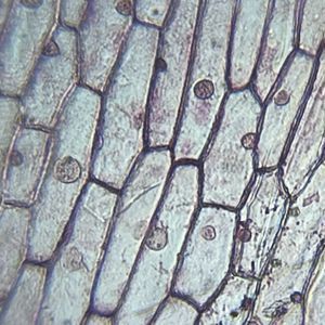
Experimenting with stains to see which gives the best view of the cell nucleus.
In order:
1. Unstained
2. Methylene Blue Stain
3. Janus Green Stain
4. Brilliant Cresyl Blue Stain
5. Bismarck Brown Stain
6. Neutral Red
All slides were wet mount slides viewed with the Foldscope (140X magnification plus the phone zoomed in to 5X magnification).
All stains were 1.0% alcohol solution (Methylene Blue was 1.0% aqueous solution) diluted to 0.1% with tap water.
Sign in to commentNobody has commented yet... Share your thoughts with the author and start the discussion!
 0 Applause
 0 Comments