LGP - Day 1 B2 : Fern Rhizome
 May 26, 2026 • 8:06 PM UTC
 India
India 340x Magnification
 Plants
Janardhan
Learn about the author...
2posts
0comments
0locations
View in Media Gallery
Hello!
My name is Janardhan KR, I'm in grade 10. I am currently attending a camp called Lodha Genius Programme at Ashoka University. I opted for "Exploring the microcosm" as my week 2 science course.
I was very excited to use a foldscope since microscopes and telesopes have always fascinated me(for example: my WhatsApp profile picture is also a picture of a phone screen under the microscope). The Foldscope 2.0 is an absolutely amazing tool, it is very intuitive to use, yet its very powerful.
We started off the session by unboxing the Foldscope. I spent a good amount of time exploring all the components of the kit. After familiarising myself with the Foldscope, I looked at the sample provided in the kit.
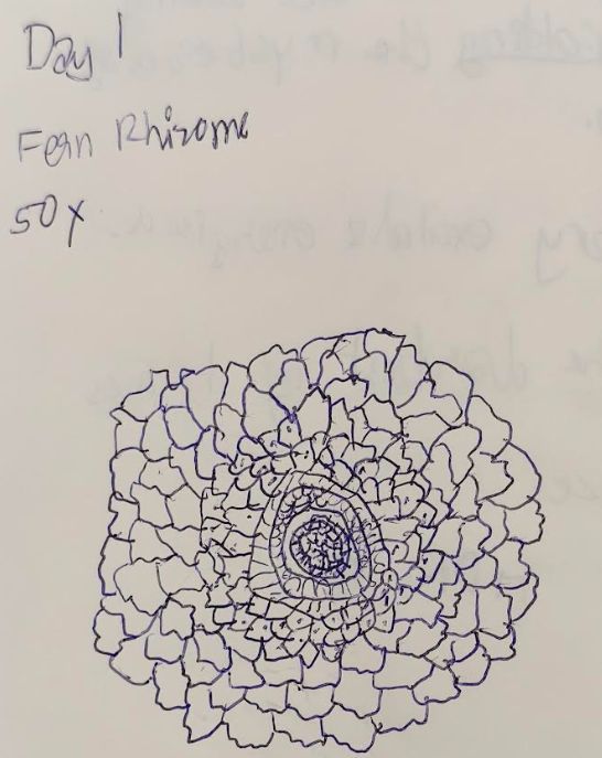
The permanent slide had a stained sample of the fern rhizome. I first looked at it under the 50x lens. I did not know what to expect, but I was mindblown to see the clarity and detail that a paper microscope could have. After looking at it carefully, I started noting down a few observations.
A few interesting things:
The sample was bursting with colour, which created a beautiful image.
I also noted that the sample had alternating bands of colour(mainly cyan and magenta).
The cells were small in the center and bigger outside .
A few pictures:
My name is Janardhan KR, I'm in grade 10. I am currently attending a camp called Lodha Genius Programme at Ashoka University. I opted for "Exploring the microcosm" as my week 2 science course.
I was very excited to use a foldscope since microscopes and telesopes have always fascinated me(for example: my WhatsApp profile picture is also a picture of a phone screen under the microscope). The Foldscope 2.0 is an absolutely amazing tool, it is very intuitive to use, yet its very powerful.
We started off the session by unboxing the Foldscope. I spent a good amount of time exploring all the components of the kit. After familiarising myself with the Foldscope, I looked at the sample provided in the kit.
The permanent slide had a stained sample of the fern rhizome. I first looked at it under the 50x lens. I did not know what to expect, but I was mindblown to see the clarity and detail that a paper microscope could have. After looking at it carefully, I started noting down a few observations.
A few interesting things:
The sample was bursting with colour, which created a beautiful image.
I also noted that the sample had alternating bands of colour(mainly cyan and magenta).
The cells were small in the center and bigger outside .
A few pictures:
Next, I switched to the 140x to get a more detailed view of the fern rhizome. I changed the lens carefully, without changing the field of view, I looked into the foldscope and I saw that I was looking at a much smaller part of the rhizome, but in much higher detail. After adjunsting the focus a little, I got a very clear image. This particular setup produced views that looked like something out of a fantasy book.
My observations:
There were 3 types of cells: The cells at the center were "dry" and very densely packed. I also noticed that some of the pink cells had a bezeled look to them(I call them crystalline(only because of their appearance)) . Finally most of the cells seemed slightly "liquidier", they seemed to have more organelles/globules present inside the cell wall and the cytoplasm seemed "more fluid" in a way.
Images:
My observations:
There were 3 types of cells: The cells at the center were "dry" and very densely packed. I also noticed that some of the pink cells had a bezeled look to them(I call them crystalline(only because of their appearance)) . Finally most of the cells seemed slightly "liquidier", they seemed to have more organelles/globules present inside the cell wall and the cytoplasm seemed "more fluid" in a way.
Images:
Finally, I could move onto the 340x lens, this would give me a very detailed view. However, when I changed the lens and gave it shot - absolutely nothing. I was very worried, but also surprised. I tried adjusting the focus, but it didn't work. I tried changing the focus very slowly and - the image popped itno focus - clear as day. This image was very unique and the amount of detail it captured was simply unbelievable. However, it was also challenging to ensure the slide remained perfectly still and the focus didn't change. Overall, it was a very fun(and a little annoying) activity to ensure everything was properly focused.
One thing I noticed was that when i was fully zoomed in, I could see a few globules in all the cells, including what I had previously called crystalline. I also had only a few cells in my field, and I really began to appreciate how so many different cell types are placed right next to each other.
Images:
One thing I noticed was that when i was fully zoomed in, I could see a few globules in all the cells, including what I had previously called crystalline. I also had only a few cells in my field, and I really began to appreciate how so many different cell types are placed right next to each other.
Images:


PS: One of the hardest challenges for me was getting the alignment and focus consistently right. Apart from that, since the slide was already prepared,I had no issue with that.
General observations:
Cell rings seemed to be arranged concentrically
About 2 -3 rings(of cells) of the same type were next to each other, after 2-3 rings, the colour and cell structure also seemed to have changed.
Getting a photo proved to be the ultimate challenge , as it needs to be constantly readjusted.
The Foldscope 2.0 is an absolutely amazing tool.
I also learnt how to start sampling better and build a good intuition for the Foldscope.
Janardhan
General observations:
Cell rings seemed to be arranged concentrically
About 2 -3 rings(of cells) of the same type were next to each other, after 2-3 rings, the colour and cell structure also seemed to have changed.
Getting a photo proved to be the ultimate challenge , as it needs to be constantly readjusted.
The Foldscope 2.0 is an absolutely amazing tool.
I also learnt how to start sampling better and build a good intuition for the Foldscope.
Janardhan
Sign in to commentNobody has commented yet... Share your thoughts with the author and start the discussion!

 0 Applause
 0 Comments