LGP26 B-2 Onion Cells - Rahul
 May 27, 2026 • 9:00 AM UTC
 India
India 340x Magnification
 Plants
Rahul
Learn about the author...
2posts
0comments
0locations
View in Media Gallery
Hello everyone,
This was my first time preparing the slide for the Foldscope. While there were many ups and downs, the overall experience and image of the onion cells were unexpected and amazing.
This was my first time preparing the slide for the Foldscope. While there were many ups and downs, the overall experience and image of the onion cells were unexpected and amazing.

View in Media Gallery
Above is a diagram of the onion cells at 50x. This diagram doesn't fully show the number of visible cells, but it does show the rigid rectangular shape of the cell due to the plant cell wall. The nuclei of the cells are somewhat visible, however the water molecules do interfere with the results.

View in Media Gallery
This is the onion sample at 140x magnification. You can see the green color of the onion cells. In additon, the nucleus are more visible. I believe the green color comes from the chlorophyll as it is a plant cell.

View in Media Gallery
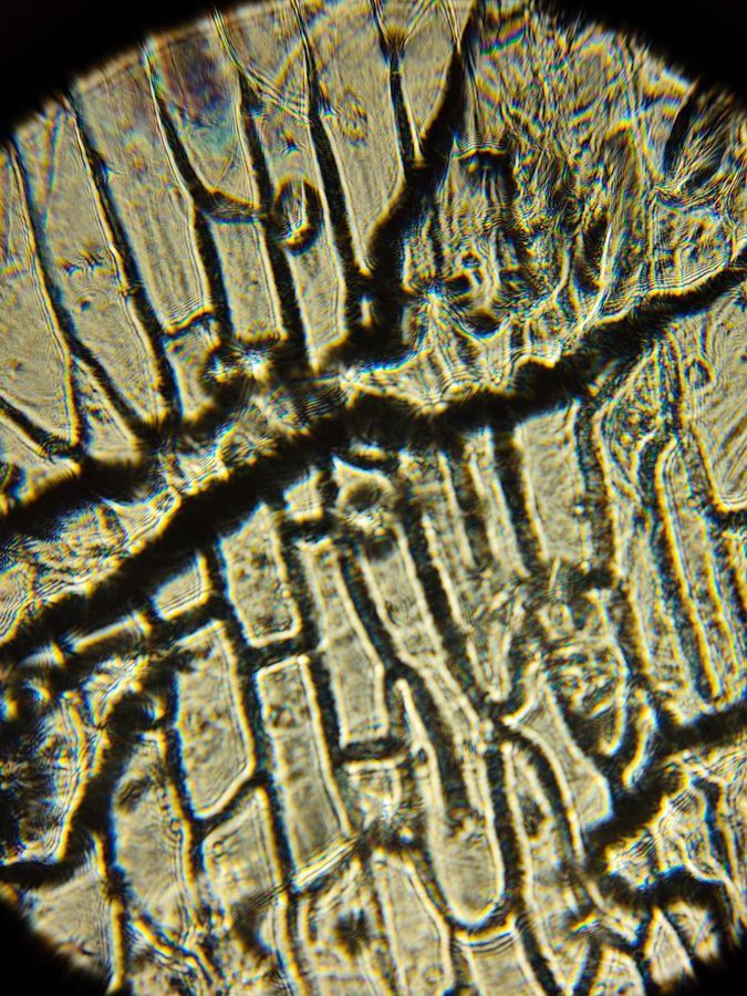
This is the 340x magnification image. As you can see, the image is slightly blurry due to poor camera quality. However, looking through the foldscope, it is very easy to see the cells and their respective components. This image offers a different perspective of an onion.
-Rahul
-Rahul
Sign in to commentNobody has commented yet... Share your thoughts with the author and start the discussion!
 0 Applause
 0 Comments