Pollen of silk floss flower
 Dec 13, 2018 • 10:58 AM UTC
 Unknown Location
 140x Magnification
 Microorganisms
Divya Kaur
Learn about the author...
11posts
0comments
1locations
View in Media Gallery
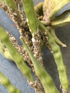
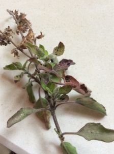
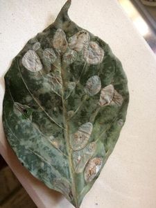
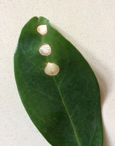
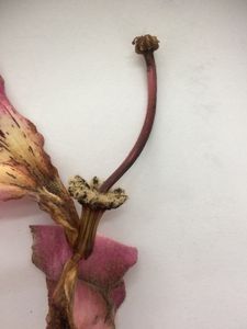
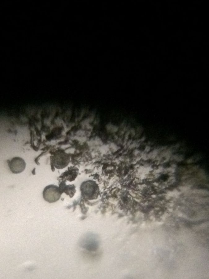
The stamens of the silk flower pollen are unique as the filaments of the five stamens fuse together forming a long dark-pink tube – red coloured staminal tube which encloses the pistil within. The anthers also join to form a canopy at the top. The anthers were scraped open to release the pollen and observed using a foldscope.
View in Media Gallery
Staminal tube
View in Media Gallery
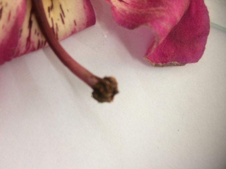
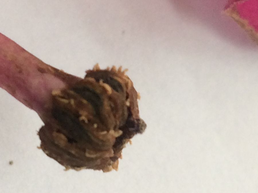
Anthers fused to form a canopy
View in Media Gallery
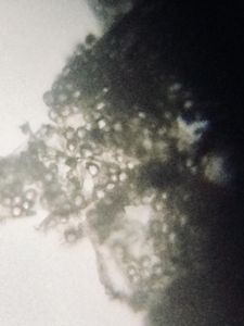
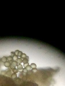
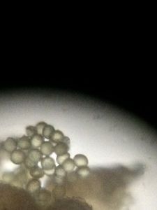
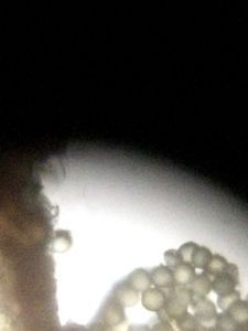
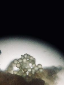
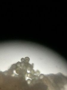
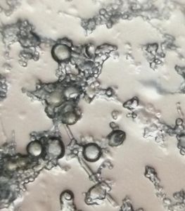
Pollen released from anther
Sign in to commentNobody has commented yet... Share your thoughts with the author and start the discussion!
 0 Applause
 0 Comments