Winter specimens at Princeton
 Jan 11, 2016 • 4:32 PM UTC
 Unknown Location
 140x Magnification
 Microorganisms
Kathleen Mulligan
Learn about the author...
1posts
0comments
1locations

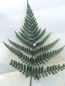
Did you know fern’s have these hard, nob-like structures all over the underside of their leaves? We didn’t either. In an effort to find something interesting to observe with the Foldscope on the Princeton campus, our group of three was looking at every leaf, rotten banana, and dead critter we could find. As we passed by the row of fern’s lining the side of Whitman dining hall, we grabbed at one and were surprised and intrigued to feel something unexpected on the underside of the leaf. This perfect combination of mystery and ecological relevance was a must-see under the Foldscope!
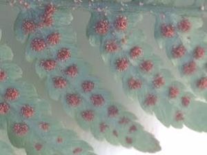
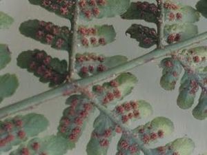
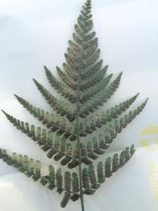
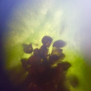
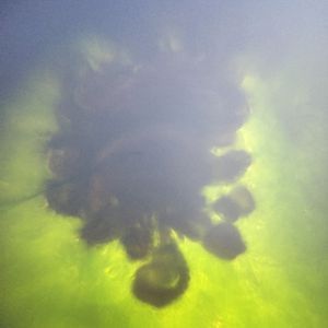
It turns out that what we were observing were the structures that encase spores. Our TA informed us that these spores are involved in the reproduction of the plant. A closer look under the Foldscope showed that each cluster contained many smaller units. After a quick search on Encyclopedia Britannica we learned that the larger clusters are called sori (singular: sorus) and the smaller units seen under the Foldscope are called sporangia. The sporangia are the spore-producing structures, each containing multiple spores. There are tons of sori on each individual leaflet, forming a dense pattern on either side of the leaf stalk, and each seem to contain many tiny sporangia.
We had many questions about fern spores. Why are there so many of them? Is a fern less apt to survive if it is deficient in these structures? What advantages are there to reproducing with spores instead of seeds? How does having spores affect their dispersal compared to other plants? The most amazing thing about the spores is how incredibly abundant they are on each fern. Every single leaf of every single fern was completely covered (we checked). Another interesting thing about these plants is that the spores are found exclusively on the bottom/underside of the leaf. Why are they all on the underside? How did this evolve? We would be curious to find out about the specific mechanisms by which these structures operate in fern plants. None of us had ever observed a plant with spores before, and the ability to see it under the Foldscope was really fascinating.
We were very excited to be able to observe something different and unusual at the macroscopic level, and take that curiosity even further with the Foldscope to the microscopic level. The three of us are all big on plants and botany, so it was a real treat to see the spores and other plants up close on our very own Princeton campus.
As a bonus, here are some of the other really interesting pictures we took from around campus!
View in Media Gallery
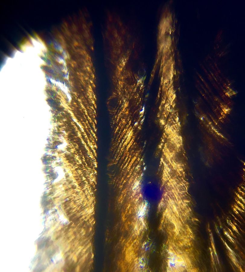
An unidentified bird feather. We were very interested in the vanes, which appeared to be fractals. We wondered to what microscopic extent they would hold the the same kind of pattern!
View in Media Gallery
This image is of some small chunks of the inside peel of a rotten banana we found nearby. Gross? Maybe, but it was for science! What level of decomposition was this in? What causes the more rotten areas to darken?
View in Media Gallery
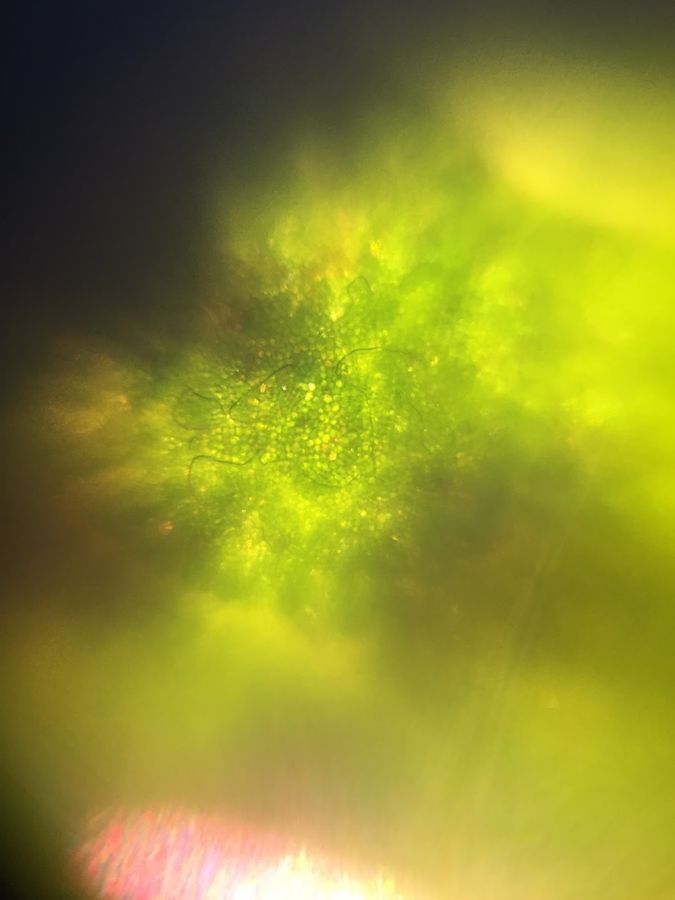
One of our first successful images captured using the Foldscope. This leaf was found near some other ones on the same bush that were growing some sort of red fungal infection – was there anything about this leaf in particular that was making it less vulnerable, or had it just not spread to this leaf yet?
View in Media Gallery
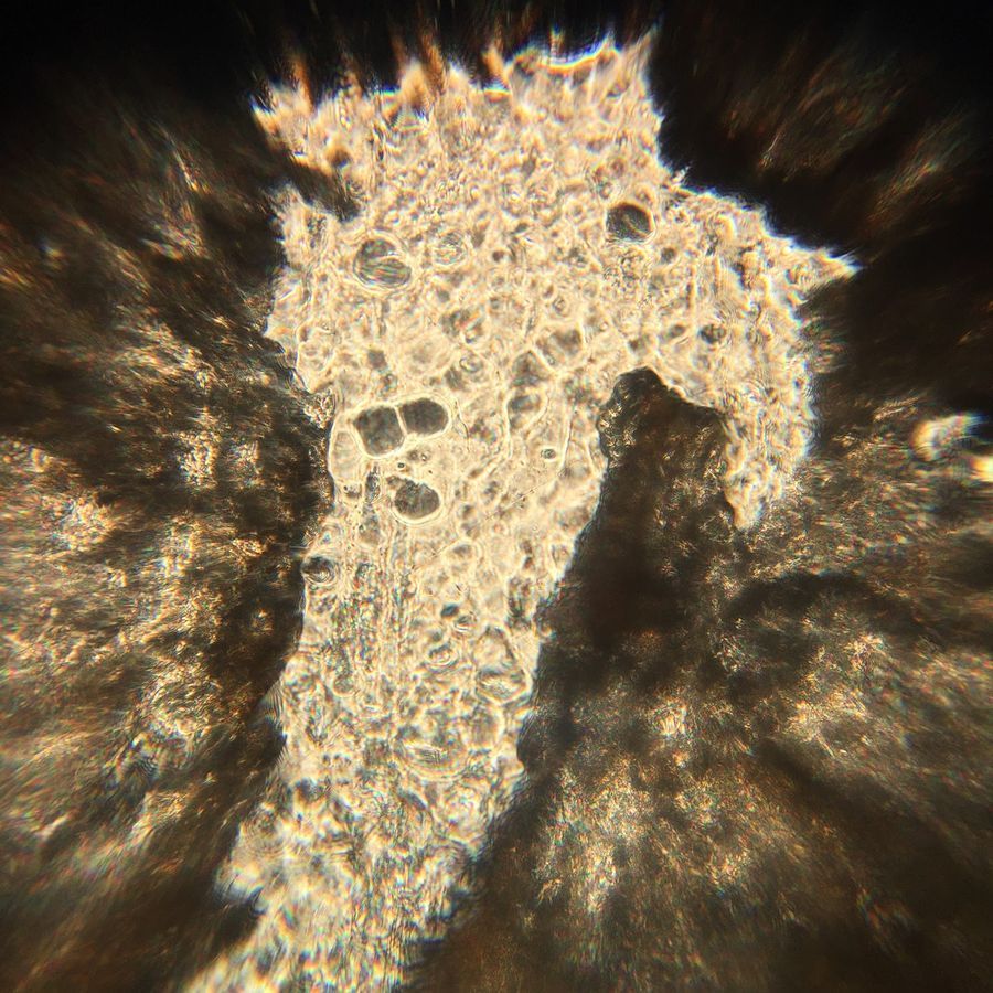
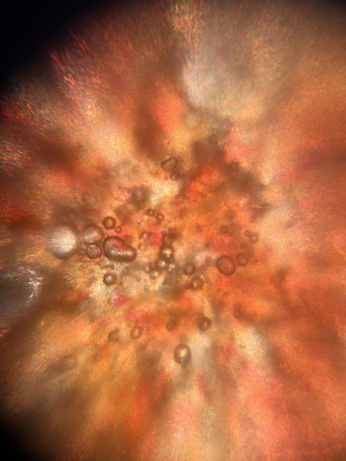
The inside of a crushed berry we collected from a tree outside the Lewis Science Library. Are those bubble-looking objects trapped air in our prepared slides, or are they from the berry itself? What accounts for all of the color variation that gives this image its water-color quality?
Jonathan, Justine and I conducted this project as part of Professor Pringle’s EEB321 class at Princeton University.
Jonathan, Justine and I conducted this project as part of Professor Pringle’s EEB321 class at Princeton University.
Sign in to commentNobody has commented yet... Share your thoughts with the author and start the discussion!
More Posts from Kathleen Mulligan
No more posts from this author.