Foot skin microbiome [Bio60_2019]
 Feb 11, 2019 • 4:03 PM UTC
 Unknown Location
 140x Magnification
 Unknown
Emma Rashes
Learn about the author...
2posts
0comments
1locations

View in Media Gallery
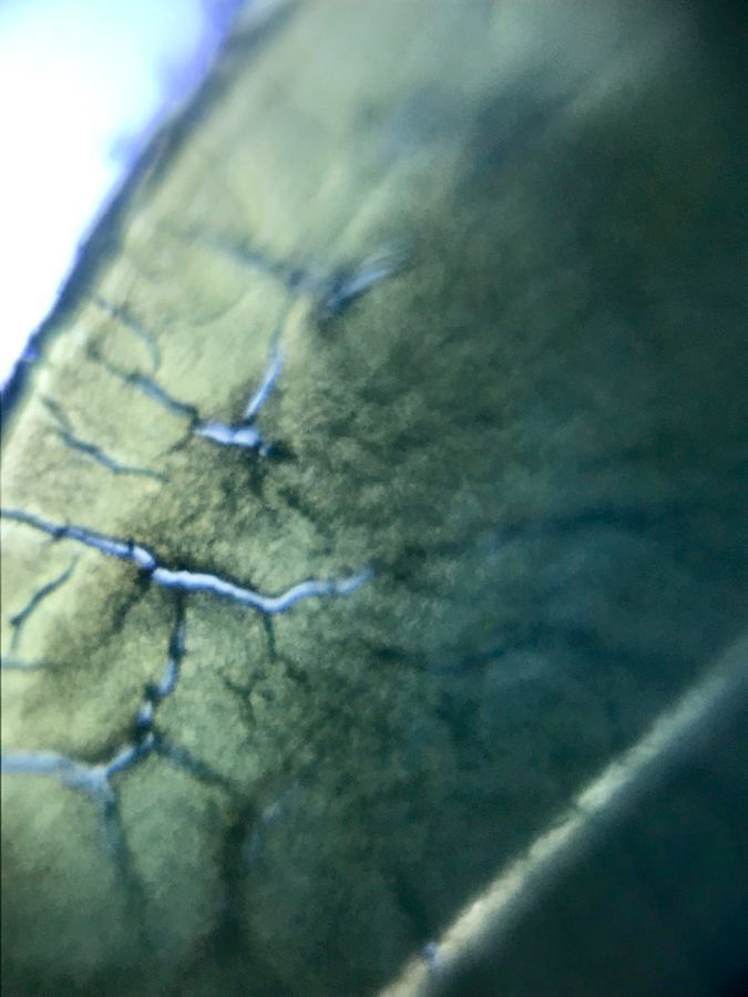
In my biology class I had the opportunity to swab and plate samples from my hands and feet, in order to explore the skin microbiome. I was interested in comparing colonies with my foldscope, as comparing colony morphology with the naked eye can be difficult. Bellow I compare two colonies that were similar in shape and size, but slightly different in color.
View in Media Gallery
Dark yellow colony immediately after making slide
View in Media Gallery
Dark yellow colony ~5 hours after making slide I photographed the dark yellow colony both immediately after creating the slide and about 5 hours later when the sample had dried onto the slide more.
View in Media Gallery
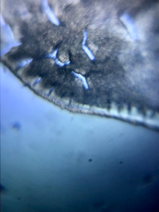
Pale yellow colony ~5 hours after making slide I photographed the pale yellow colony about 5 hours after making the slide. The pale yellow colony seems less dense. The pale yellow colony also has a less distinct color even under the foldscope.

View in Media Gallery
Plate that bacteria were collected from
Sign in to commentNobody has commented yet... Share your thoughts with the author and start the discussion!
![Thumbnail for Dead skin- [Bio60_2019]](https://storage.googleapis.com/microcosmosdelta.appspot.com/images/transfer_129425/IMG_6890_300x300.jpeg)
 0 Applause
 0 Comments