Microscopic explorations in a Jo’burg garden
 Feb 03, 2016 • 9:12 AM UTC
 Unknown Location
 140x Magnification
 Unknown
Dorit Hockman
Learn about the author...
8posts
40comments
1locations

I have just arrived back in South Africa from the UK. While spending the weekend visiting my parents in Jo’burg, I thought I’d take the opportunity to show them the microscopic wonders they may not have been aware of lurking in their very own garden.

My parents’ sunny Jo’burg garden A tiny caterpillar (which I suspect was a very young larva of a geometer moth, also known as an inch-worm, due to its distinctive crawl) happened to inch its way across my arm, and I immediately went for my Foldcsope. This first image shows how tiny it was compared to the Foldcsope ruler, probably about 1.5mm in length. Under the scope, using the high mignification lens, iPhone attachment and LED light module, we could clearly see some surprisingly beautiful details on this tiny caterpillar. I was especially impressed by the beautiful spots that accompanied each tiny sensory hair along the length of the body (see “inch_worm” movie below).

Tiny inch worm
Inspired by the inch-worm, I went for a wonder around the garden to collect a menagerie of flora and fauna for my next slide. The image below shows the slide displaying a fruit fly I stole from an old spider’s web, a young aphid from a hibiscus plant, a piece of the stigma (top of the pollen tube) from the bright red flower of the same plant and a small piece of fern frond, including a single sorus or spore bundle.

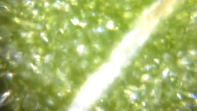
A selection of flora and fauna I focused on the fern frond first, and was intrigued by the striped pattern detailing the edge of each individual sporangium, where the spores are found in the sorus (shown below in the “Fern-frond” movie). The bright green cells of the leaf bordered by pure white veins, resembled a stained glass window pierced by rays of bright sunlight . The veins at the front edge were strikingly similar in arrangement to those I’d seen on the leading edge of a bee’s wing, during my first Foldscope adventure . In the image below the movie, you can see them side-by-side. When I mentioned this to my mom, a mathematician, she immediately started pondering the mathematical principles that likely underlie the development of this striking example of convergent evolution.


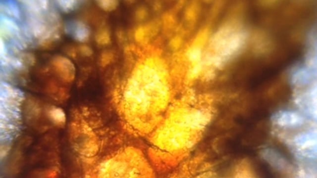
Comparing the branching veins of a bee’s wing to those in a fern frond Moving onto the aphid, the colours of this tiny arthropod (which I slightly squished, so is surrounded by yellow liquid in the “aphid” movie below) were astounding: the beautiful burnt orange of its abdomen were in striking contrast to the completely translucent, ghostly appearance of its legs. So too was the bright magenta of the hibiscus stamen, shown below the aphid movie.


Hibiscus stamen Finally, I moved on to the fruit fly, which had probably been decaying for while in the spider’s web. The wings were still in tact so, I focused on the leading edge of the wing and discovered what I suspect must be a broad patch of sensory bristles at the leading edge of the wing. I wonder how the anatomy this patch varies in different species? I will have to try find some more flies!
Watch this space for more microscopic adventures from Cape Town, my next destination!
Watch this space for more microscopic adventures from Cape Town, my next destination!

The leading edge of a fly’s wing
Sign in to commentNobody has commented yet... Share your thoughts with the author and start the discussion!

 0 Applause
 0 Comments


