Prostatic hyperplasia
 Apr 20, 2019 • 3:11 AM UTC
 Unknown Location
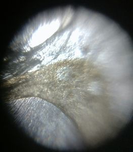
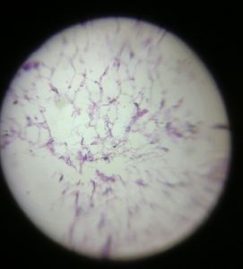
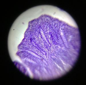
 140x Magnification
 Unknown
RDGMC Ujjain 4 4
Learn about the author...
54posts
1comments
1locations

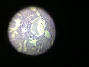
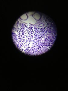
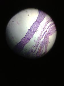
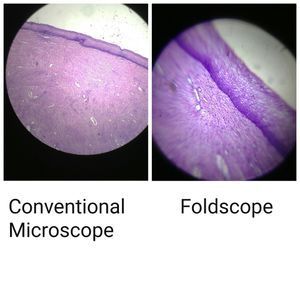
Section shows normal prostatic tissue with back to back arrangement of glands with columnar epithelium as seen under Foldscope.
Sign in to commentNobody has commented yet... Share your thoughts with the author and start the discussion!
 0 Applause
 0 Comments