Microcosmos Logo
ExploreAll PostsAboutShop Now
ExploreAll PostsAbout
Log In or Sign Up

Salivary Gland

Applause IconAug 24, 2018 • 12:11 AM UTC
Location IconUnknown Location
Applause Icon140x Magnification
Applause IconMicroorganisms
RDGMC Ujjain 4

Learn about the author...

54posts
1comments
1locations
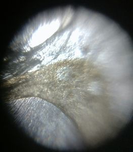
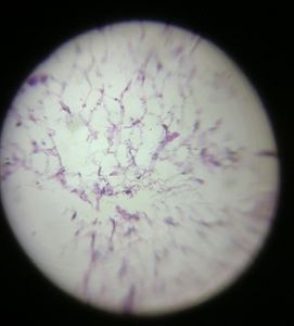
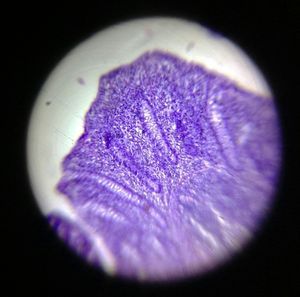
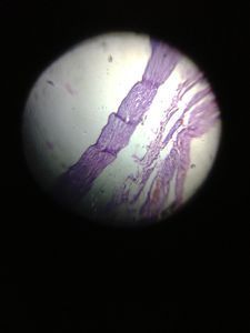
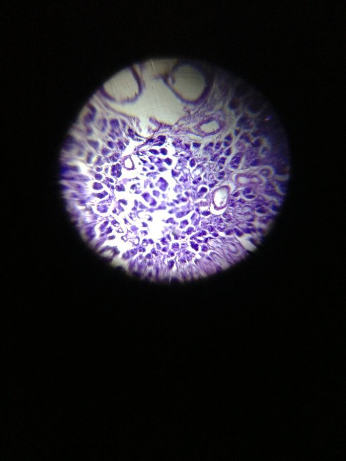
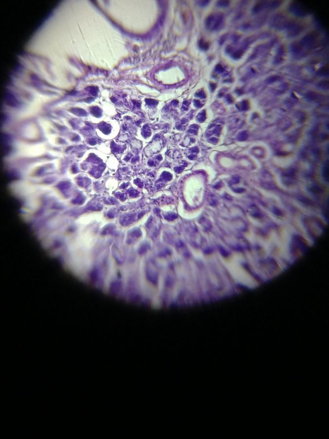
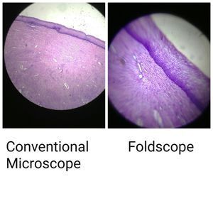
The section shows normal salivary gland tissue including glands in tubules formation as seen under Foldscope from Department of Paediatrics and the Department of Pathology of R D Gardi Medical College, Ujjain

Sign in to commentNobody has commented yet... Share your thoughts with the author and start the discussion!

More Posts from RDGMC Ujjain 4

Comparision between Foldscope and Conventional microscope
0 Applause0 Comments
6y
Lipoma
0 Applause0 Comments
6y
Prostatic hyperplasia
0 Applause0 Comments
6y
Ascaris under “Foldscope”
0 Applause0 Comments
6y
Old skin of snake
0 Applause0 Comments
6y
Foldscope Utility Workshop
0 Applause0 Comments
6y
Chronic Appendicitis
0 Applause0 Comments
6y
Lipoma
0 Applause0 Comments
6y
Chronic Appendicitis
0 Applause0 Comments
6y
Epithelial Hyperplasia
0 Applause0 Comments
6y
Fallopian Tube
0 Applause0 Comments
6y
Cholecystitis
0 Applause0 Comments
6y
Squamous cell carcinoma
0 Applause0 Comments
6y
Abdominal Tuberculosis
0 Applause0 Comments
6y
Gastric Mucosal Atrophy
0 Applause0 Comments
6y
Ovarian Cyst
0 Applause0 Comments
6y
Inflammatory Lesion
0 Applause0 Comments
6y
Features suggestive of lipoma
0 Applause0 Comments
6y
Small Intestine
0 Applause0 Comments
6y
Endometrium
0 Applause0 Comments
6y
Microcosmos

About

Explore

All Posts

Brought to you by

Foldscope

© 2024 Microcosmos

Contact

Terms and Conditions

Social Media