The flowerful foldscope: Flowers and Fruit on Stanford Campus
 May 09, 2019 • 11:10 PM UTC
 Unknown Location
 140x Magnification
 Fungi
Fungi
Mihyun Choi
Learn about the author...
2posts
0comments
1locations


We got a white flower from a tree on Stanford campus. Its petal and anther were imaged using the Foldscope. All photos were taken with a Samsung Galaxy S8. The scale bar was obtained by imaging a caliper set to 0.2mm, obtaining the distance between the calipers on the image, then creating a 100um scale bar that is half that distance. The size of the field of view was calibrated to the caliper photo’s field of view.

View in Media Gallery
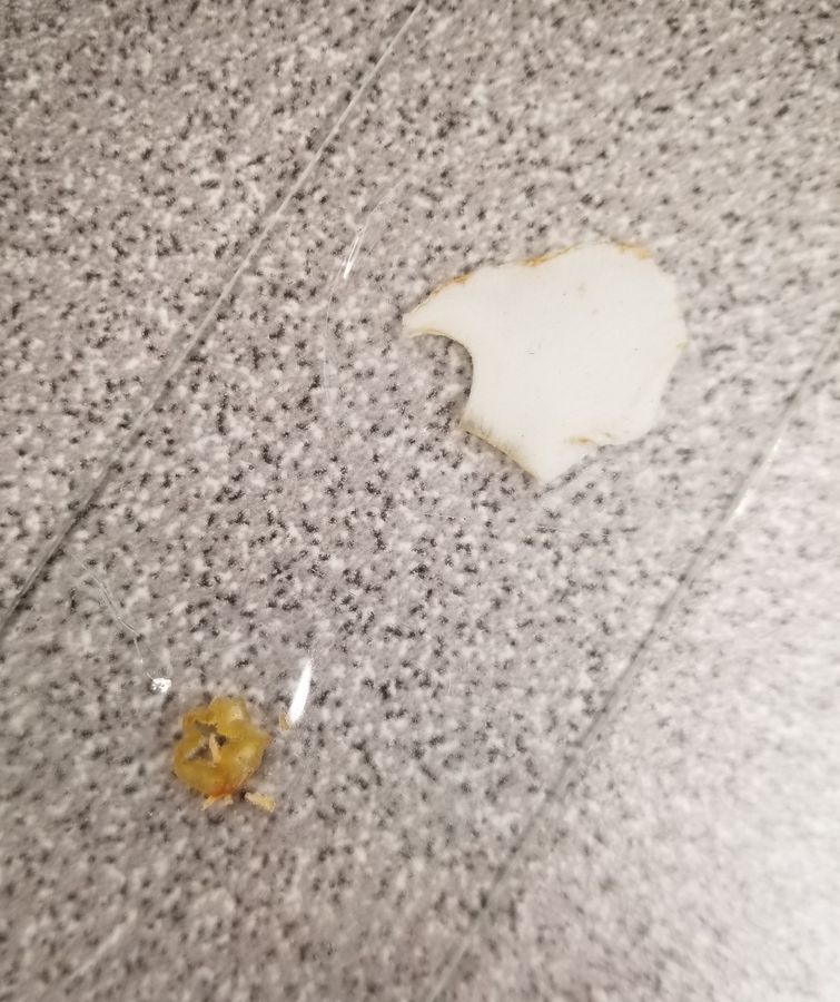
This is a picture of the petal. To the naked eye, the petals look like there aren’t any patterns on them. However, under the Foldscope, we could see the petal veins. Also, there were a few pollen particles on them shown in yellow.

View in Media Gallery
This is an image of the anther under the Foldscope. The anther is the pollen-bearing body of the flower. Less light passes through the anther compared to the petal, meaning that it is relatively compact. There are small hair-like extensions on the anther.
View in Media Gallery
A bigger white flower was also examined. From this flower, the petal and the bottom of the flower, where the ovaries and ovules reside in the pistil, were studied.

View in Media Gallery
Under the Foldscope, the granules in this petal was finer than the smaller flower before, even tho the flower itself was bigger. They have a shiny crystal-like visual.

View in Media Gallery
The pistil was of a similar density as the anther. It was very liquidy. No finer pattern was on the surface of the pistil.

We also collected a petal from a bigger pink flower to see if there is a difference in the microstructure based on the color.
View in Media Gallery
We could visualize the microstructures of the petal using the Foldscope. It had an organization to it where there were circular subunits, which could be pigments or cells.

View in Media Gallery
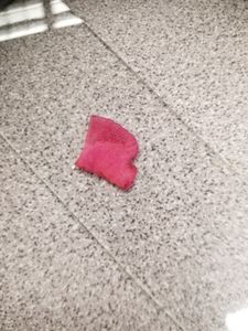
We also found red berries on a small tree. A berry was collected, cut in half, and pressed against the glass slide, so that the skin is facing upward.

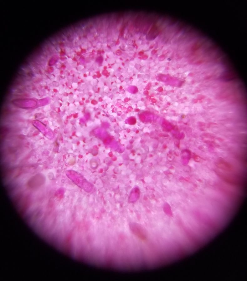
It was harder to focus on this globular structure, but the skin had small irregular microstructures in them. We think it looks like the sun.
Please let us know if you can identify the species of any of the above.
Please let us know if you can identify the species of any of the above.
Sign in to commentNobody has commented yet... Share your thoughts with the author and start the discussion!
 0 Applause
 0 Comments