I can’t be-leaf it!!! A survey of the leaves on Stanford campus
 May 10, 2019 • 12:58 AM UTC
 Unknown Location
 140x Magnification
 Microorganisms
Mihyun Choi
Learn about the author...
2posts
0comments
1locations
View in Media Gallery
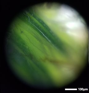
We observed various types of leaves we could find on campus using the Foldscope. Pictures were taken with the Samsung Galaxy S8. The scale bar was obtained by imaging a caliper set to 0.2mm, obtaining the distance between the calipers on the image, then creating a 100um scale bar that is half that distance. The size of the field of view was calibrated to the caliper photo’s field of view.

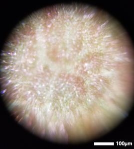
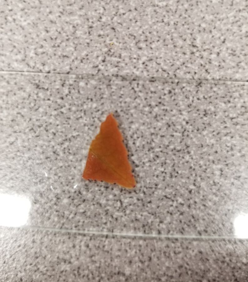
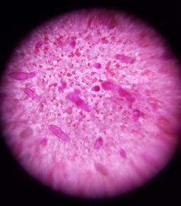
We picked up a brown leaf, but observing the leaf macroscopically (left), there are shades of green. We decided that it would be interesting to see if this green pigment was cell specific; we wanted to see if cells were green, brown, or both. Using the foldscope (left), we can see the cell patterning of the leaf. Interestingly, there are spots of red and green within each cell! Some parasitic plants may contain red pigment, because they do not photosynthesize, meaning that they don’t need green chlorophyll. Carotenoids and anthocyanins may provide yellow, orange, red, or purple coloring shown in this leaf.
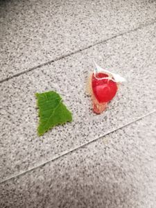


We stumbled upon another leaf that had an interesting edge pattern (left). This leaf had ridges that looked like it’d poke you, but observing the edges closer (center), we see that they are actually rounded. Indeed, when touching the leaf, it doesn’t actually poke you, and it feels soft! Looking at the edge, we noticed an interesting clear branching vein pattern. Looking towards the center of the leaf (right), we could see that these veins are laid throughout the entire leaf.


Outside of the building in which we work, we found a tree that had semi-pointy leaves. These leaves are characteristic to conifers. Through the fold scope, we found that there weren’t many veins that we found on the other leaves which we might expect. Given their slender nature, perhaps they do not need as an extensive branching network as a normal leaf.
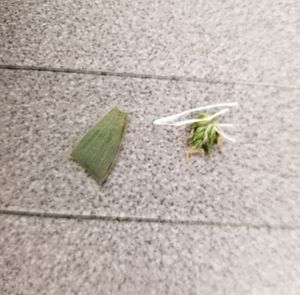

Next, we observed a leaf with white hairs (center) and a flower bud (right). The leaf is interesting, because there were silvery/white hairs, which can even be seen microscopically. The flower bud has bright green areas with a dark green center.


Our last two leaves had interesting vein patterns. In the middle, we can see narrow clear veins, while on the right, there are veins that end in the middle of the chlorophyll segments. We put these two images side by side, because microscopically, we see a simple leaf with veins, but through the foldscope, we see that they actually have different vein patterns!
We strongly encourage you to go outside and see if you can find/identify these leaves we have shown!
We strongly encourage you to go outside and see if you can find/identify these leaves we have shown!
Sign in to commentNobody has commented yet... Share your thoughts with the author and start the discussion!
 0 Applause
 0 Comments