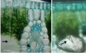
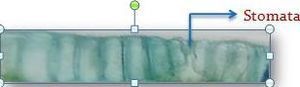
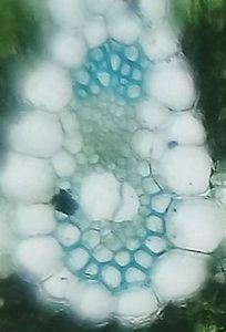
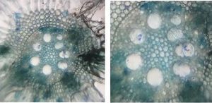
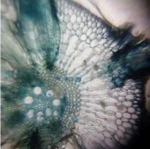
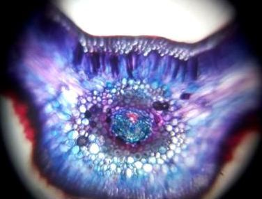
T.S of Cycas leaflet stained with safranin and fast green
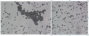
 May 21, 2019 • 5:54 AM UTC
 Unknown Location
 140x Magnification
 Microorganisms
SOUMYA P.R
Learn about the author...
30posts
0comments
1locations
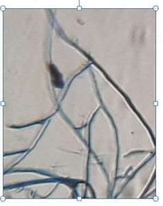
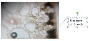
View in Media Gallery
The image shows the epidermis, hypodermis and vascular bundle region. Upper epidermis surrounded by a layer of cuticle. Below the upper epidermis, sclerenchymatous hypodermis region is present. Mesophyll is differentiated into palisade and spongy parenchyma. A single vascular bundle present, showing xylem and phloem.
Sign in to commentNobody has commented yet... Share your thoughts with the author and start the discussion!

 0 Applause
 0 Comments