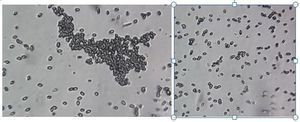
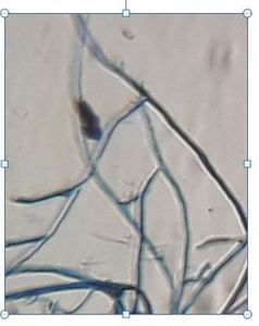
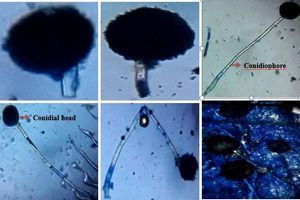
Penicillium sp. stained with Lactophenol cotton blue

Applause IconMay 25, 2019 • 8:12 AM UTC
Location IconUnknown Location
Applause Icon140x Magnification
Applause IconMicroorganisms

Learn about the author...

30posts
0comments
0locations
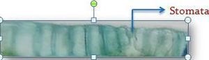
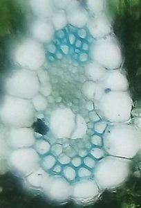
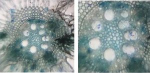
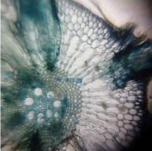
The microscopic structure of Penicillium sp. stained with lactophenol cotton blue shows the conidiophores and conidia. Conidia (spores) are produced from the tips of the phialids. The fungi were isolated from polluted lake water.

Sign in to commentNobody has commented yet... Share your thoughts with the author and start the discussion!

More Posts from SOUMYA P.R

Penicillium sp. stained with Tryphan Blue
0 Applause0 Comments
5y
Fungal spore stained with Tryphan Blue
0 Applause0 Comments
5y
Fungal spore stained with Lactophenol cotton blue
0 Applause0 Comments
5y
Fungal spore stained with methylene blue
0 Applause0 Comments
5y
Microscopic structure of fungal hyphae stained with Tryphan blue
0 Applause0 Comments
5y
Fungal hyphae stained with Methylene blue
0 Applause0 Comments
5y
Fungal hyphae stained with lactophenol cotton blue
0 Applause0 Comments
5y
Presence of sclereids in the petiole of E. crassipes – Stained with Tryphan blue
0 Applause0 Comments
5y
Histochemical study for the presence of starch in the petiole of E. crassipes
0 Applause0 Comments
5y
Presence of sclereids in the leaf of E. crassipes
0 Applause0 Comments
5y
Enlarged portion of epidermis and stomata
0 Applause0 Comments
5y
Enlarged portion of vascular bundle – T.S. of petiole (E. crassipes) – Stained with Safranin
0 Applause0 Comments
5y
Enlarged portion of vascular bundle -T.S. of petiole (Eichhornia crassipes) – Stained with Tryphan blue
0 Applause0 Comments
5y
Enlarged portion of vascular bundle -T.S. of leaf (E. crassipes)
0 Applause0 Comments
5y
T.S. of root of E. crassipes – enlarged portion of vascular bundle
0 Applause0 Comments
5y
T.S. of root of Eichhornia crassipes stained with methylene blue.
0 Applause0 Comments
5y
T.S of Cycas leaflet stained with safranin and fast green
0 Applause0 Comments
5y
Fungal spore: Microscopic structure of A. niger stained with tryphan blue
0 Applause0 Comments
5y
T.S. of leaf (E. crassipes) stained with methylene blue.
0 Applause0 Comments
5y
T.S. of Petiole (E. crassipes) stained with tryphan blue
0 Applause0 Comments
5y