Various parts from Ladybug
 May 21, 2019 • 11:23 PM UTC
 Unknown Location
 140x Magnification
 Unknown
Vincent Tieu
Learn about the author...
5posts
0comments
2locations
View in Media Gallery
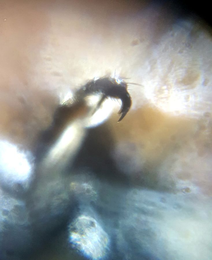
A dead ladybug was found in my room on May 10th, 2019 at 5:00 pm. Instead of letting a good opportunity go to waste, I decided to dissect it and image its parts under the Foldscope lens. The above picture is of one of its back legs. I removed the leg and wet-mounted it onto a glass slide. You can see the tibia and claw of the leg in this picture, as well as some hairs, which possibly act as feel receptors.
View in Media Gallery
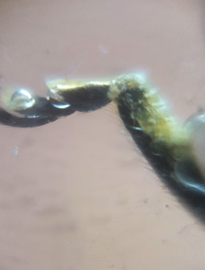
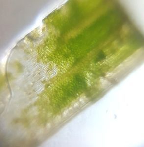
This is a picture of a mandible from the same ladybug. The head from the ladybug was removed via razor blade, the mandible was then sliced off, and finally mounted onto a glass slide. You can see the claw structure, which will help with eating, and the hairs which act as feelers.
View in Media Gallery
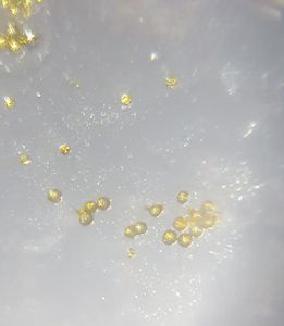
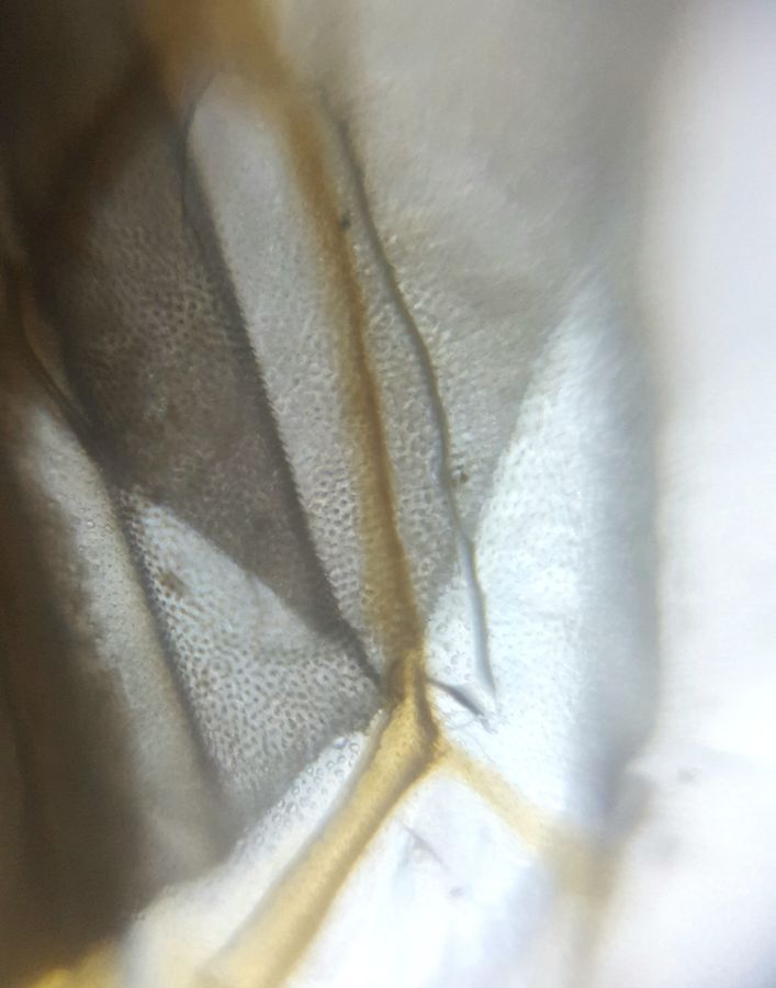
This is a picture of the wings of the ladybug. I removed the hard protective shell to reveal the delicate wings folded underneath. I then peeled off a wing and wet-mounted it to a glass slide. The brown structure running from the top to bottom in the picture is likely a vein providing support to the wing. The black spots are most likely the tiny hairs covering the entire membrane of the wing.
View in Media Gallery
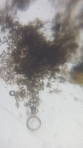
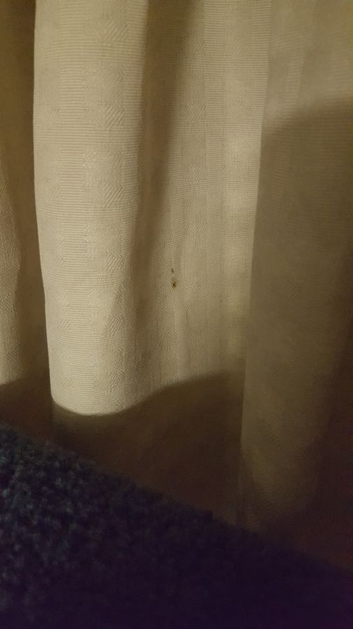
All of the pictures are of the same ladybug, which was found dead in the curtains in my room, on May 10th, 2019, 5:00 pm.
#caltechbi1
#caltechbi1
Sign in to commentNobody has commented yet... Share your thoughts with the author and start the discussion!
 0 Applause
 0 Comments