Close encounters with an amoebic kind
 Jun 29, 2016 • 8:21 AM UTC
 Unknown Location
 140x Magnification
 Microorganisms
Laks Iyer
Human observer of life. https://sukshmadarshin.wordpress.com
97posts
1255comments
5locations
View in Media Gallery
Over the summer I have decided to work with one or a few micronauts at a time every weekend. This time I thought I’d let them join me in my explorations. Most importantly I want to share that observations aren’t always picture perfect and ideas aren’t always good to start with, but as you keep observing, you refine your observations and soon you might have something exciting at hand. I have had two such sessions this far; the previous one with Nataraj, Yash and Kartik, and this one was with Reethi. These one-on-one sessions are really satisfying. I am posting our observations in reverse order as I cannot contain my excitement about what emerged in the observation session with Reethi.
The water sample is the same from two weeks ago ( see https://microcosmos.foldscope.com/?p=16915 ). By last week, it had a terrible odor of H2S and while we saw a bunch of Daphnia suggesting that the sample wasnt completely anaerobic, there were a good number of ciliates in it (Future post). Last Sunday (after ~ two weeks), the odor was gone, but yet there seemed to be a lot of activity. We used the Ditch-spacer slide technique to view these.
The following videos were taken towards the end of the session with Reethi. We saw this object which we thought might be an amoeba. We set up the camera and returned 22 minutes later. What a surprise!
1.
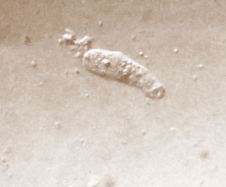
Figure 1. Amoeba-17130-1-LI
I wasn’t very pleased with the lighting and so I turned to my table lamp as a light source and kept searching until I found this single type of amoeba that was widely present in the slide. This one looked different from the previous one. I have over 30 minutes of footage and so I had the chance to study different aspects of the amoeba (temp id: Amoeba-17130-2-LN). Firstly, here is a compilation of its motion. The following video is run 4x faster.
2.
Figure 2. Amoeba-17130-2-LN
I also have several videos of it eating/interacting with bacteria and possibly phagocytosis snapping. Then there is a flagellate that is very commonly seen in the slide and I have a strong suspicion it is the flagellate stage of the same amoeba.
I also have several videos of it eating/interacting with bacteria and possibly phagocytosis snapping. Then there is a flagellate that is very commonly seen in the slide and I have a strong suspicion it is the flagellate stage of the same amoeba.
Figure 3. Feeding dynamics and possible flagellate stage
Based on this my strongest suspicions are that this is a Vahlkampfid heterolobosean amoeba, like Naegleria or a related species. Vahlkampfia like organisms are also called amoeboflagellates. These are very common in pond water.
The interesting thing is that amoebae are not monophyletic, which means that all amoebas are not necessarily close to each other. Vahlkampfid protists are actually a sister group of kinetoplastids ( Leishmania, Trypanosoma ) in the evolutionary tree, as compared to Amoeba proteus that are closely related to slime molds. Then there is Capsaspora , an amoeba found in the snail Biomphalaria , and is the sister group of animals and choanoflagellates. Thus finding out the exact genus and species of an amoeba is tricky. It would require isolating the organism and studying it carefully, and of course sequencing informative pieces of its DNA.
Amoebae have fascinated me from my childhood days. Cant wait to see the amoeba that is almost visible to the naked eye. With respect to this sample, I of course am exerting caution and soon plan to bleach the bottle out and discard it, but wait there is more …
Laks (with Reethi).
Sign in to commentNobody has commented yet... Share your thoughts with the author and start the discussion!

 0 Applause
 0 Comments
_300x300.jpeg)


