Cell Viability of Animal Cell Line
 Sep 18, 2019 • 11:05 PM UTC
 Unknown Location
 140x Magnification
 Unknown
Vanishree Rathod
Learn about the author...
2posts
0comments
1locations
View in Media Gallery
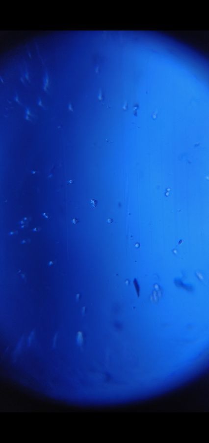
SKBR-3, a breast cancer cell was prepared on a hemocytometer and observed under Foldscope. Trypan blue staining was done and both viable and dead cells can be seen.
Procedure:
(1) Place 100 µl of cell
suspension in a cryo-vial.
(2) Add equal parts of 0.4%
trypan blue dye to the cell suspension to obtain a 1 to 2 dilution (example: 50
µl of cells to 50 µl of trypan blue) and mix by pipetting up and down.
(3) With the cover slip already
in place, fill one side of a hemacytometer counter with the cell suspension by
placing the tip of the pipette at the notch.
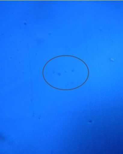
Blue cells are the nonviable
cells.
Procedure:
(1) Place 100 µl of cell
suspension in a cryo-vial.
(2) Add equal parts of 0.4%
trypan blue dye to the cell suspension to obtain a 1 to 2 dilution (example: 50
µl of cells to 50 µl of trypan blue) and mix by pipetting up and down.
(3) With the cover slip already
in place, fill one side of a hemacytometer counter with the cell suspension by
placing the tip of the pipette at the notch.
Blue cells are the nonviable
cells.
View in Media Gallery
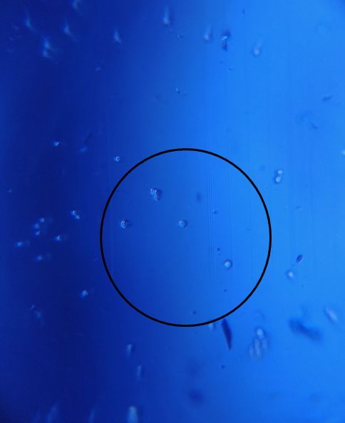
Viable cells as seen under Foldscope
View in Media Gallery
Non-viable Animal cells under Foldscope
Sign in to commentNobody has commented yet... Share your thoughts with the author and start the discussion!
 0 Applause
 0 Comments