Mouse Pancreas for Histological Analysis
 Sep 18, 2019 • 11:59 PM UTC
 Unknown Location
 140x Magnification
 Microorganisms
Vanishree Rathod
Learn about the author...
2posts
0comments
1locations
View in Media Gallery
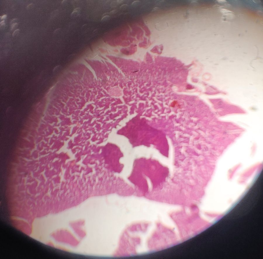
Thin sections of mouse pancreas were stained by H&E and observed under Foldscope and Microscope.
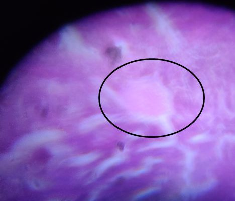
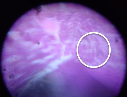
Beta cells and Islet endothelial cell can be seen with Foldscope.
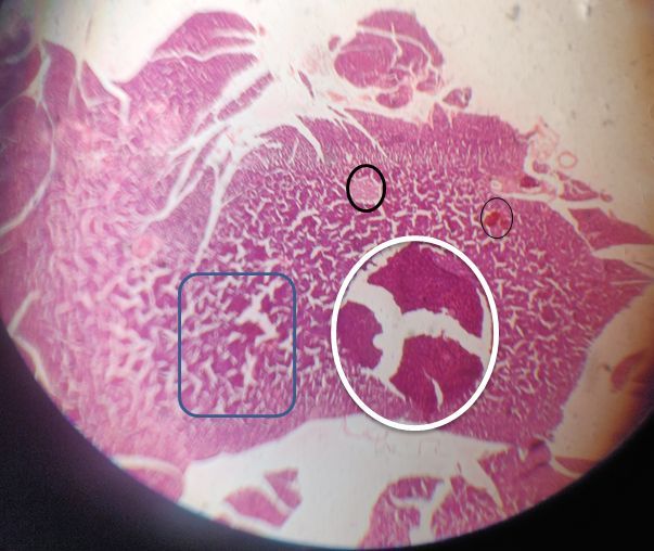
Beta cells, Islet endothelial cell , Pancreatic acini and interlobular duct can be seen under compond microscope.
Pancreatic acini is stained darker and has a fibrous wall.
Beta cells and Islet endothelial cell can be seen with Foldscope.
Beta cells, Islet endothelial cell , Pancreatic acini and interlobular duct can be seen under compond microscope.
Pancreatic acini is stained darker and has a fibrous wall.
View in Media Gallery
Beta cells under Foldscope
View in Media Gallery
Islet endothelial cell
under Foldscope
under Foldscope
View in Media Gallery
Under microscope beta cells ( bold black circle),
Pancreatic acini (black circle), interlobular duct ( white circle) and
Islet endothelial cell ( blue box) can be observed.
Pancreatic acini (black circle), interlobular duct ( white circle) and
Islet endothelial cell ( blue box) can be observed.
Sign in to commentNobody has commented yet... Share your thoughts with the author and start the discussion!

 0 Applause
 0 Comments