Foldscope photography: my journey from simply observing to clicking high quality photos.
 Nov 18, 2020 • 4:21 AM UTC
 Unknown Location
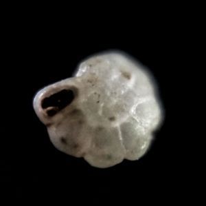
 140x Magnification
 Unknown
Dr Rafikh Shaikh
I'm a science educator interested in everything science.
115posts
92comments
20locations
View in Media Gallery
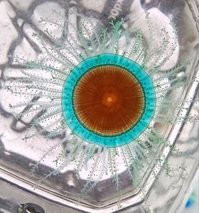
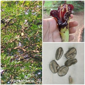
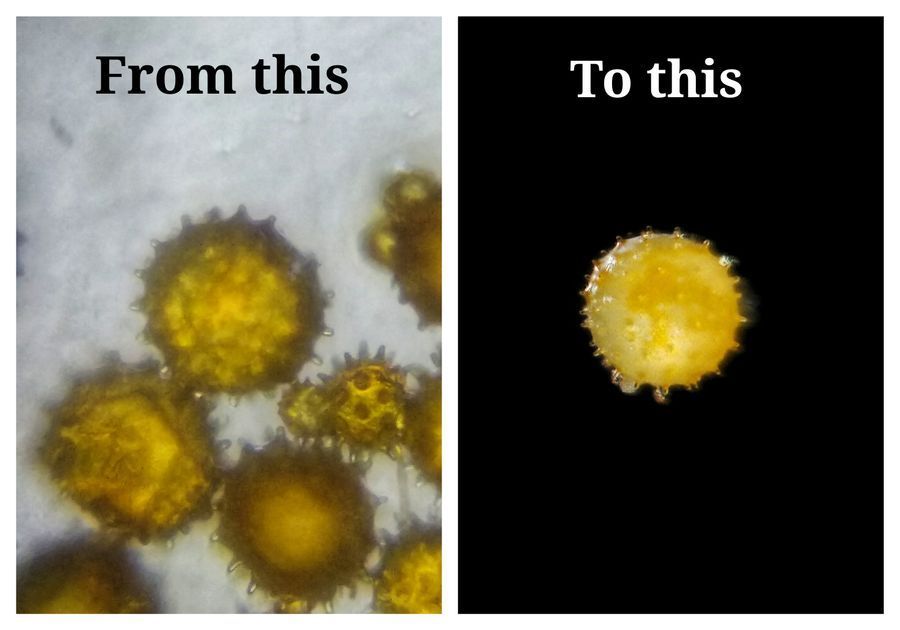
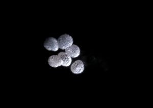
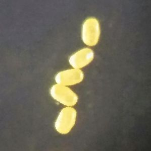
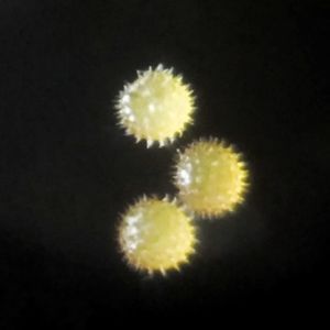
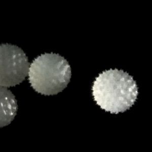
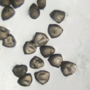
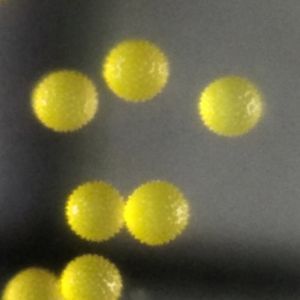
Pollens of hibiscus flower taken with two different techniques We all use Foldscope to observe microscopic world and occasionally take photos to share with community. I have been doing it from last few years but then I thought can I take high quality photos with Foldscope, photos so good that they can be submitted to microscopic photography competitions.
To answer my questions I have been experimenting with Foldscope. I tried various lighting arrangments, slide preparation methods, editing techniques. In this post I am listing out some tips which work:
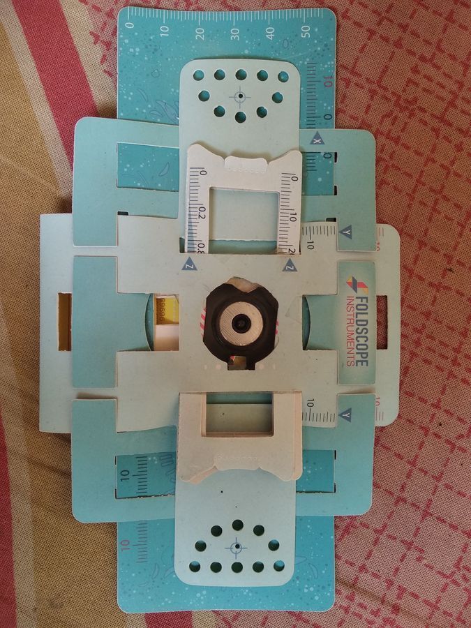
Lighting arrangments: for many samples such as pollens shinning light from top works best. I have two holes (as shown in image) near lens from where I shine light, one is for phone flashlight and other is for a tiny LED light. Shinning light from top has advantage it brings out the true colours of the object.
To answer my questions I have been experimenting with Foldscope. I tried various lighting arrangments, slide preparation methods, editing techniques. In this post I am listing out some tips which work:
Lighting arrangments: for many samples such as pollens shinning light from top works best. I have two holes (as shown in image) near lens from where I shine light, one is for phone flashlight and other is for a tiny LED light. Shinning light from top has advantage it brings out the true colours of the object.
View in Media Gallery
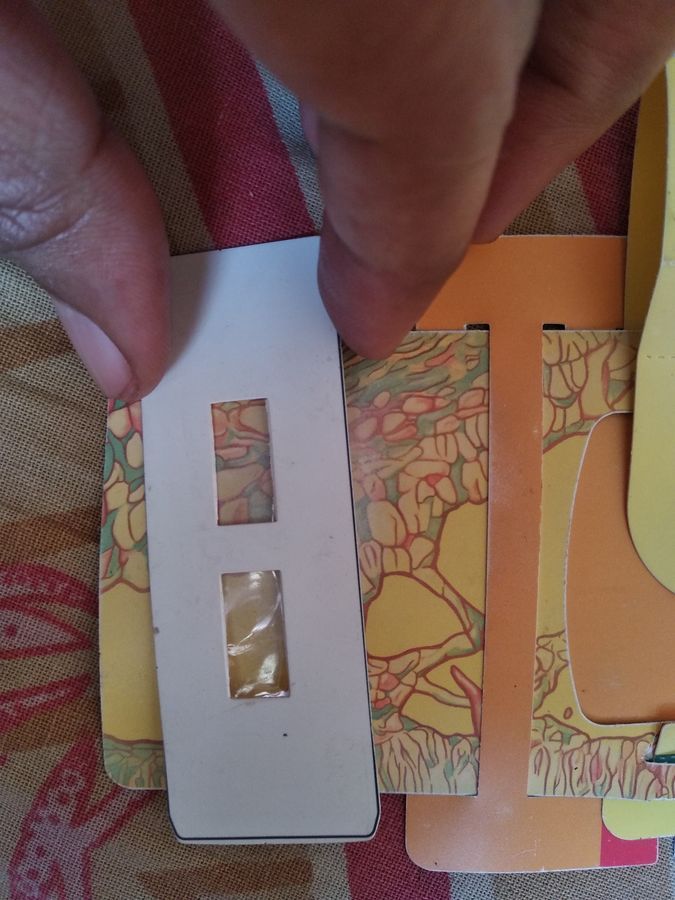
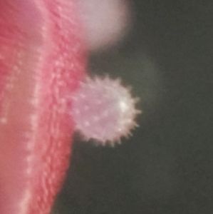
Image showing two holes near the lens for shining light. 2. Slide preparation: many samples are safe to be viewed without covering them with tape or coverslips. For pollens I use the paper slide and put tape from just one side so pollens do fall. While observing I put uncovered side towards the lens. Covering with tape deforms the object you are observing, for example compound eyes of insects or pollens, they get stuck to sticky side of tape and looks weird.
View in Media Gallery
Image showing slide with one side covered with tape 3. Editing: in many cases when object is big and it can’t be focused in one photo, I take multiple photos and focus stack them. I use an app called Snapseed on phone and GIMP on laptop. Here is a detailed post by @manup on how to focus stack images.
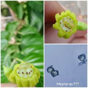
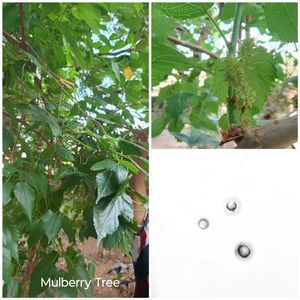
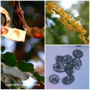
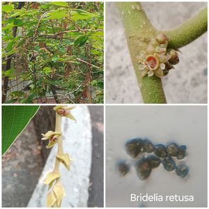
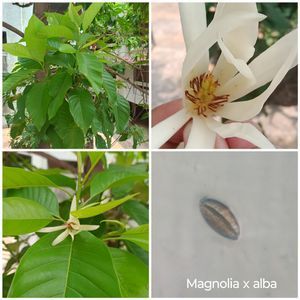
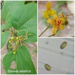
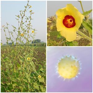
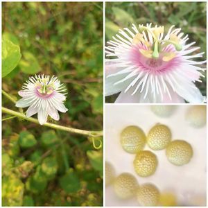
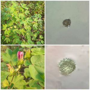
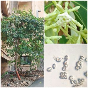
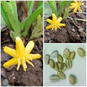
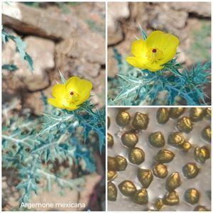
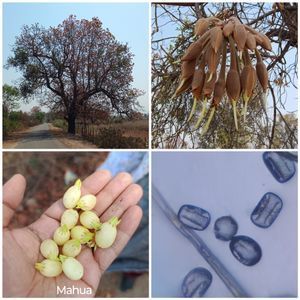
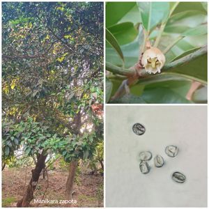
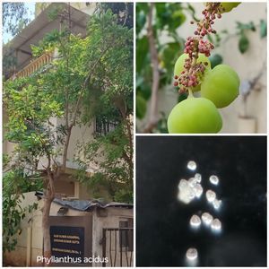
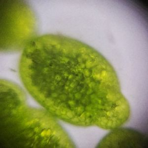
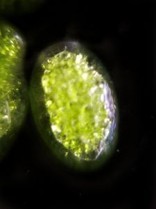
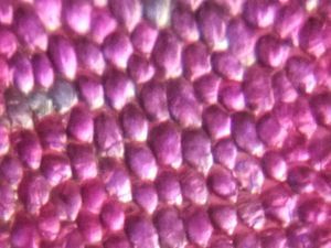
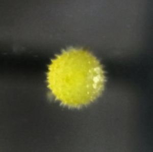
Foldscope focus stacking for high depth of field images That’s it, that is all you need to get amazing photos with Foldscope. Here are some pictures I am proud of (I have submitted 3 photos from these to a competition):
Foldscope focus stacking for high depth of field images That’s it, that is all you need to get amazing photos with Foldscope. Here are some pictures I am proud of (I have submitted 3 photos from these to a competition):
Let me know your thoughts on this post and sharing your experiences with microscopic photography. Best, Edurafi.
Sign in to commentNobody has commented yet... Share your thoughts with the author and start the discussion!

 0 Applause
 0 Comments