Blood prepared slides testing
 Mar 05, 2015 • 6:08 PM UTC
 Unknown Location
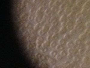
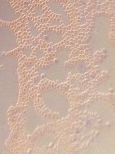
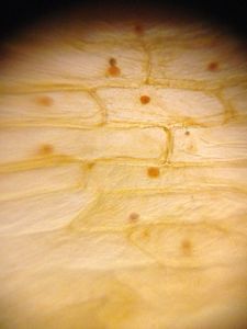
 140x Magnification
 Microorganisms
Don Meunier
Learn about the author...
9posts
0comments
1locations
View in Media Gallery
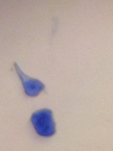
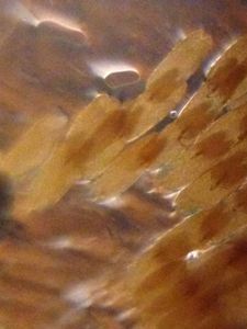
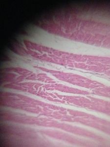
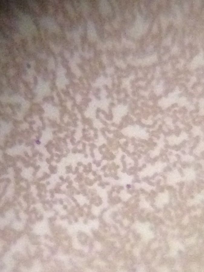
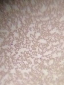
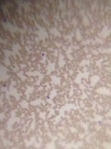
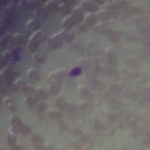
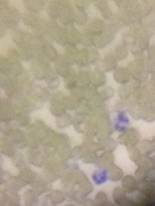
I am very interested in seeing what I can do with the Foldscope, examining blood smears. Here are prepared slides, Wright’s stain. 2 low mag and 1 high mag. I am having difficulty with getting a good image with the high mag lens. The LED light gives a cast around the cells. I will try my blood next without cover slips. With digital zoom, the WBC’s are clearly distinguished.
Sign in to commentNobody has commented yet... Share your thoughts with the author and start the discussion!
 0 Applause
 0 Comments