Captured rat skin section by Foldscope
 Apr 18, 2018 • 10:54 AM UTC
 Unknown Location
 140x Magnification
 Microorganisms
Jyotchna Gogoi
Learn about the author...
10posts
2comments
1locations
View in Media Gallery
I was really very anxious to see my histology slides after attending the Foldscope workshop organised in ICGEB by DBT on 16/04/2018. I am happy as I could focus the section with Foldscope and mobile, which I couldn’t do in the workshop. pressing the sample stage folds in mobile phone view, section get nicely focused. But, while viewing directly, stage folds need to raised for focusing.
Thank you for inventing such handy instrument which could be used by every child.
Thank you for inventing such handy instrument which could be used by every child.
View in Media Gallery
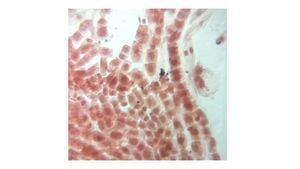
Rat skin tissue section 1
View in Media Gallery
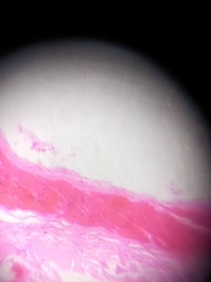
Subcutis layer

View in Media Gallery
Blood vessels

View in Media Gallery
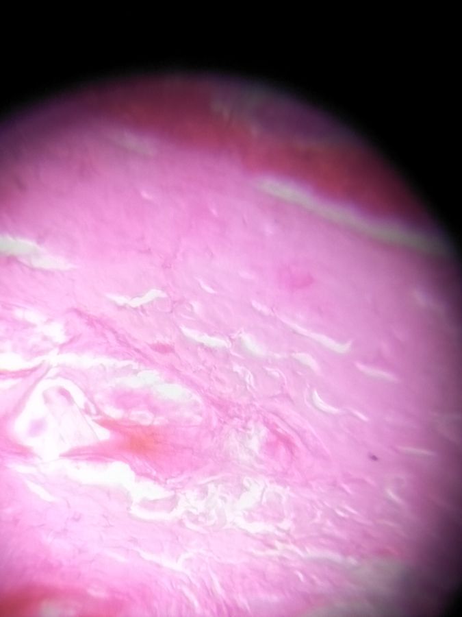
Dermis layer focus area 1

View in Media Gallery
Dermis layer focus area 2
Sign in to commentNobody has commented yet... Share your thoughts with the author and start the discussion!
 0 Applause
 0 Comments