Africa, human blood smear (ID: 4)
 May 24, 2018 • 10:53 PM UTC
 Unknown Location
 140x Magnification
 Microorganisms
Marissa Cucinotta
Learn about the author...
2posts
0comments
1locations
View in Media Gallery
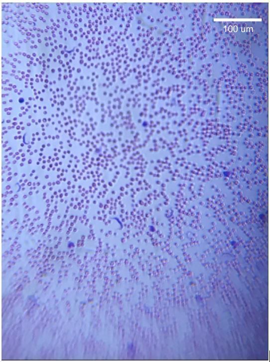
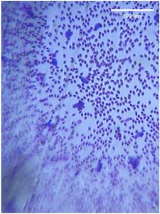
Stained human blood smear from Africa showing African Trypanosomiasis A prepared slide of a human blood smear from the African continent was provided for examination with the foldscope. The picture was taken using a smartphone with zoom. Within the blood smear, several unexpected cells were visible. They were irregularly shaped and stained darker than the red blood cells. They were long, thin, and curled into arcs. They also appeared to have flagella.
An online search on parasites in the blood revealed that the defining features of the cells aligned with African Trypanosomiasis, also known as “sleeping sickness.” Though there were a few cells visible when scanning through the smear, the parasites were not abundant in the sample. There were about one or two parasites in the field of view at any given time.
The scale bar was added by referencing the diameter of red blood cells, which is 7.3 um on average (McCormick 1927). The diameter (D) of a single red blood cell was measured in ImageJ (in units of pixels), and a scale bar was drawn for 100 um by using the ratio (D pixels) / (7.3 um). The length of the scale bar in pixels was thus calculated as 100 um * (D pixels) / (7.3 um).
Sign in to commentNobody has commented yet... Share your thoughts with the author and start the discussion!
 0 Applause
 0 Comments