US, hawk/owl (ID: 6)
 May 24, 2018 • 10:59 PM UTC
 Unknown Location
 140x Magnification
 Microorganisms
Marissa Cucinotta
Learn about the author...
2posts
0comments
1locations
View in Media Gallery
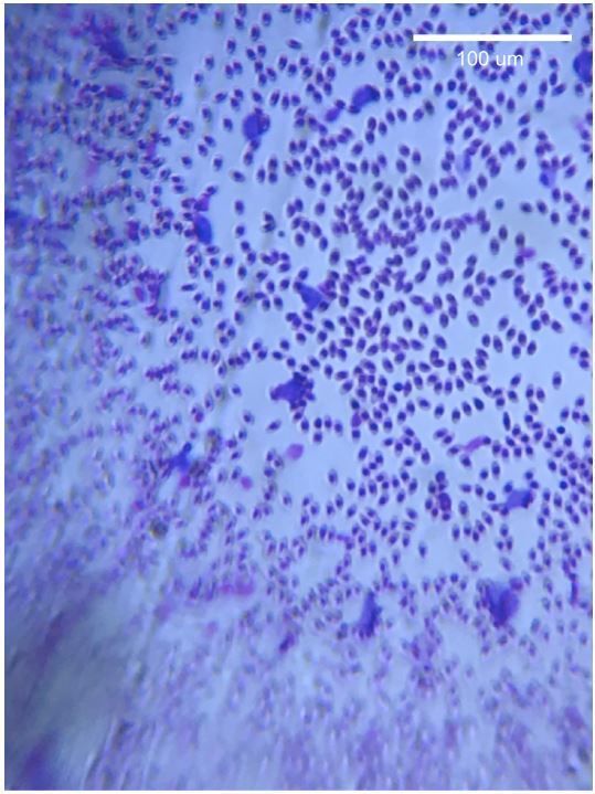
Stained blood smear from an owl or hawk in the US (ID: 6) A prepared blood smear slide from a owl or hawk in the US was provided for examination with the foldscope. The picture was taken using a smartphone with zoom. Within the blood smear, there were several red blood cells with greatly enlarged nuclei.
An internet search on avian blood diseases revealed the appearance of the enlarged cells aligned with Leucocytozoon gametocytes. The parasitic infection seemed very prevalent in the sample, with 10s of cells visible in the field of view throughout the sample.
The scale bar was added by referencing the diameter of red blood cells, which is 7.3 um on average (McCormick 1927). The diameter (D) of a single uninfected red blood cell was measured in ImageJ (in units of pixels), and a scale bar was drawn for 100 um by using the ratio (D pixels) / (7.3 um). The length of the scale bar in pixels was thus calculated as 100 um * (D pixels) / (7.3 um).
Sign in to commentNobody has commented yet... Share your thoughts with the author and start the discussion!
 0 Applause
 0 Comments