MORPHOANATOMICAL CHARACTERS OF MEDICINAL PLANTS IN MANNAR REGION
 Jun 05, 2018 • 4:26 AM UTC
 Unknown Location
 140x Magnification
 Microorganisms
S SHARMILA
Learn about the author...
9posts
4comments
1locations
View in Media Gallery
Foldscope is so affordable and can easy to used anywhere, it brings science to our daily life activities. I’m looking forward to work with the foldscope it is very easy to carry and handling a foldsope is innovative learning to me.
With the help of foldscope I observed anatomical characters in plant stem and also studied the trichome morphology. This experience was amazing to me.
Anatomy – 1 Acalypha indica Linn.
My first anatomical section was taken from Acalypha indica Linn. commonly called as Indian copperleaf belongs to the family Euphorbiaceae
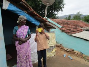
Habit of Acalypha indica
With the help of foldscope I observed anatomical characters in plant stem and also studied the trichome morphology. This experience was amazing to me.
Anatomy – 1 Acalypha indica Linn.
My first anatomical section was taken from Acalypha indica Linn. commonly called as Indian copperleaf belongs to the family Euphorbiaceae
Habit of Acalypha indica
View in Media Gallery
Here is the first foldscope image….
View in Media Gallery
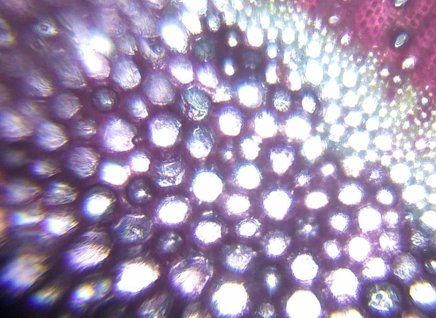
T.S of Acalypha indica stem
View in Media Gallery
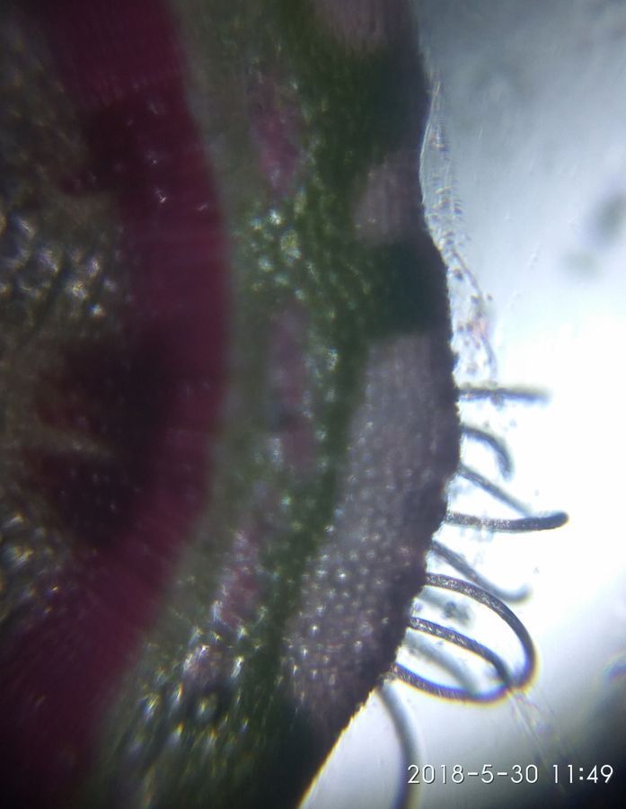
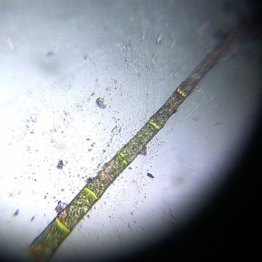
Beautiful view of Unicellular trichome
View in Media Gallery
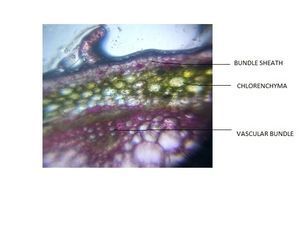
View of Vascular bundles Important characters:
Presence of thick cuticle and non-glandular unicellular trichomes in epidermal region. Followed by 2-3 layers of collenchyma and rest are parenchymatous cortex. With the help of foldscope I have clearly observed the continuous ring of vascular tissues made up of xylem and phloem. With the support of foldscope under high magnification I clearly viewed Sphaeraphides and glandular matter in cortex and pith region.
Anatomy – 2 Adhatoda vasica Nees .

View in Media Gallery
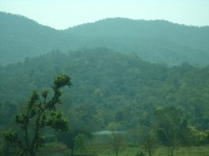
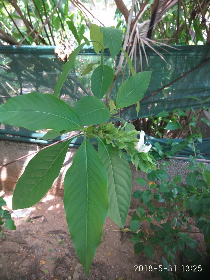
Habit of Adhatoda vasica Nees
View in Media Gallery
I went side to the tree and got a good look at that thin low branches that hold flowers
View in Media Gallery
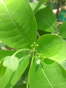
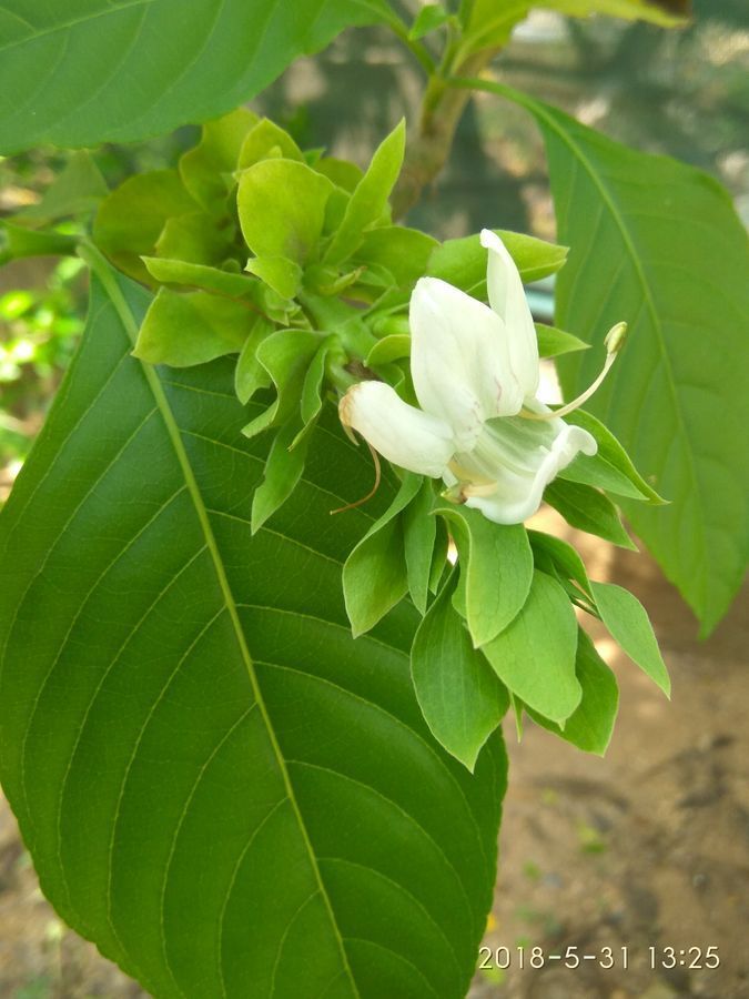
Blooming twig Adhatoda vasica commonly called as Malabar nut belongs to the family Acanthaceae.
View in Media Gallery
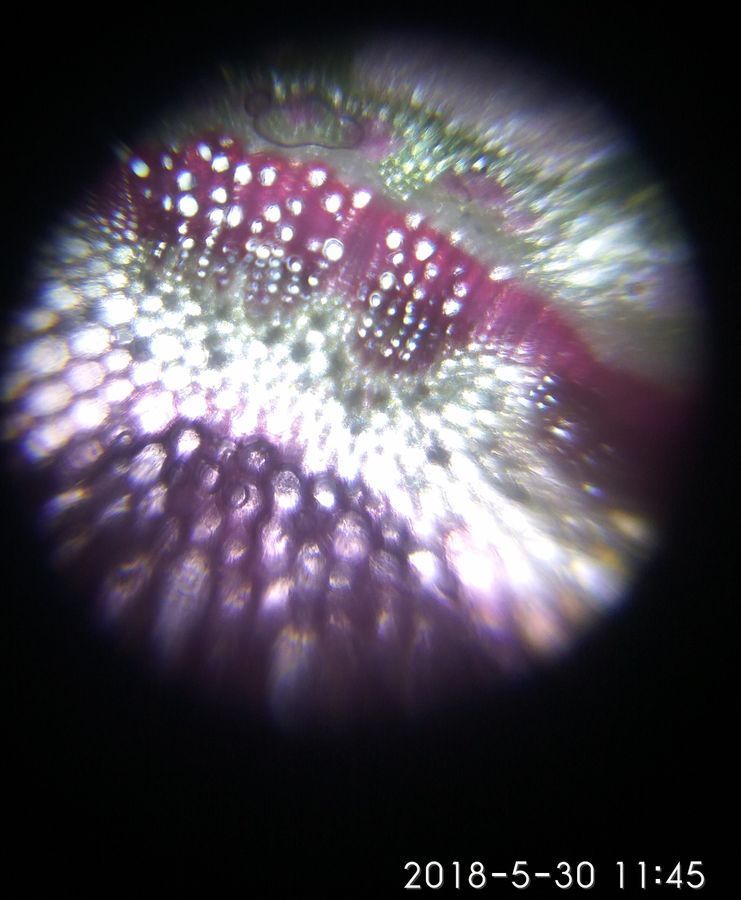
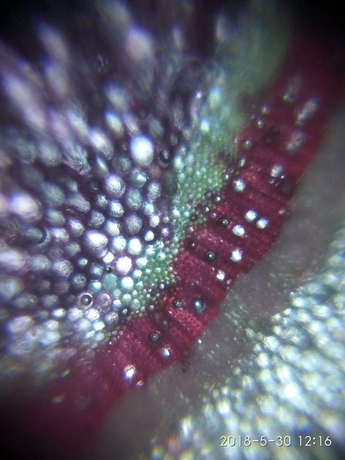
T.S of Adhatoda vasica stem – Foldscope image
View in Media Gallery
Important characters:
I adjusted focus ramp in foldscope I clearly seen the vascular bundle they are arranged in siphonostele pattern which encloses a wide central pith. Starch grains and calcium oxalate crystals are seen in the middle region under high zoom foldscope image. Euphorbia hirta L.
I adjusted focus ramp in foldscope I clearly seen the vascular bundle they are arranged in siphonostele pattern which encloses a wide central pith. Starch grains and calcium oxalate crystals are seen in the middle region under high zoom foldscope image. Euphorbia hirta L.

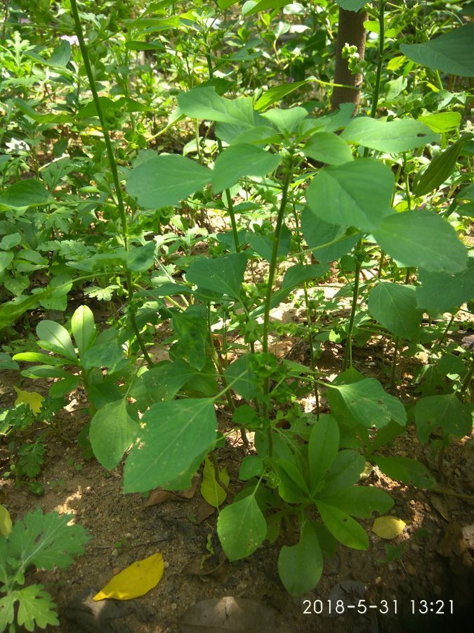
View in Media Gallery
Aerial view of Euphorbia hirta Trichomes:
View in Media Gallery
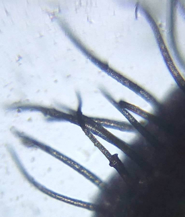
This is my eminent pic in foldscope I have never taken before – Trichome I really surprised to view the single trichome in the outer region which is multicellular and uniseriate.
At that time I awe to capture that adorable pic in my mobile phone with the help of foldscope.
So I greatfully acknowledge Dr. Manu prakash Ms. Christine Q Kurinara, and the prakash lab at Stanford university for encouraging and supporting me in this project.
I also thank department of biotechnology for providing me the wonderful opportunity to exposure myself in foldscope.
Dr. S. SHARMILA (required) drsharmilas@yahoo.com (required) Website Give me more suggestions about plant anatomy foldscope pics... Submit
Sign in to commentNobody has commented yet... Share your thoughts with the author and start the discussion!
 0 Applause
 0 Comments