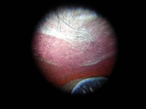
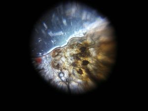
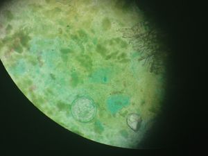
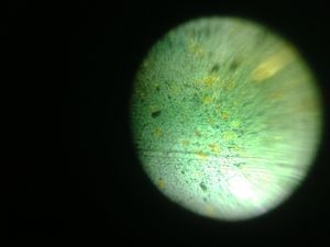
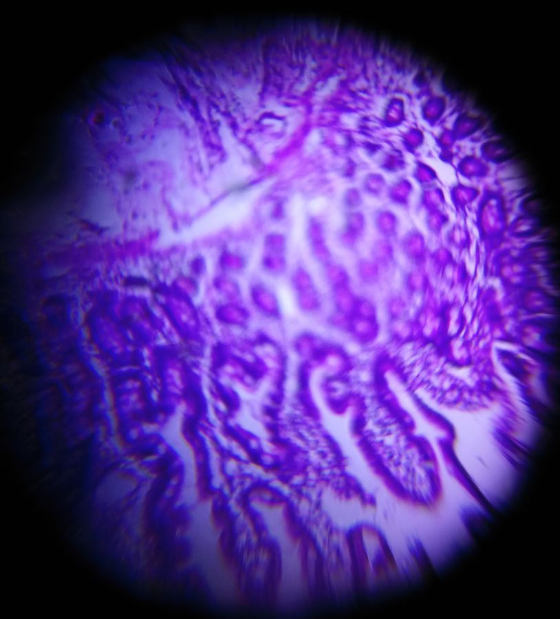
Duodenal Tissue

Applause IconJun 14, 2018 • 3:44 AM UTC
Location IconUnknown Location
Applause Icon140x Magnification
Applause IconMicroorganisms

Learn about the author...

62posts
0comments
0locations
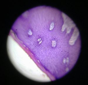
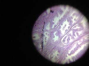
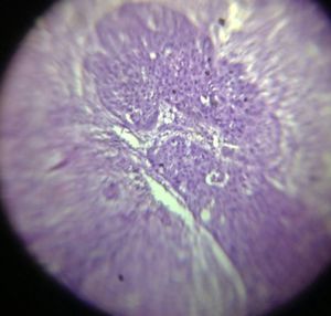
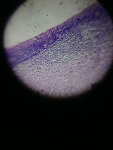
Section shows pieces of normal duodenal tissue showing villi and glands seen in a foldscope from Department of pathology of R D Gardi Medical College, Ujjain

Sign in to commentNobody has commented yet... Share your thoughts with the author and start the discussion!

More Posts from RDGMC Ujjain 6

Lipoma

Applause Icon 0 ApplauseComment Icon 0 Comments
5y
Cervix

Applause Icon 0 ApplauseComment Icon 0 Comments
5y
Sebaceous Cyst

Applause Icon 0 ApplauseComment Icon 0 Comments
5y
Blood of mosquito

Applause Icon 0 ApplauseComment Icon 0 Comments
5y
Mosquito under Foldscope

Applause Icon 0 ApplauseComment Icon 0 Comments
5y
Compound eye of mosquito under “Foldscope”

Applause Icon 0 ApplauseComment Icon 0 Comments
5y
Intestine

Applause Icon 0 ApplauseComment Icon 0 Comments
6y
Lipoma

Applause Icon 0 ApplauseComment Icon 0 Comments
6y
Ova of stool under Foldscope

Applause Icon 0 ApplauseComment Icon 0 Comments
6y
Stool sample

Applause Icon 0 ApplauseComment Icon 0 Comments
6y
Stool sample

Applause Icon 0 ApplauseComment Icon 0 Comments
6y
Workshop for “Foldscope”

Applause Icon 0 ApplauseComment Icon 0 Comments
6y
Prostatic Hyperplasia

Applause Icon 0 ApplauseComment Icon 0 Comments
6y
Carcinoma In-Situ

Applause Icon 0 ApplauseComment Icon 0 Comments
6y
Fibroadenoma

Applause Icon 0 ApplauseComment Icon 0 Comments
6y
Cervical features are consistent with prolapse

Applause Icon 0 ApplauseComment Icon 0 Comments
6y
Prostatic hyperplasia

Applause Icon 0 ApplauseComment Icon 0 Comments
6y
Metastatic lymph node

Applause Icon 0 ApplauseComment Icon 0 Comments
6y
Cervix

Applause Icon 0 ApplauseComment Icon 0 Comments
6y
Ovarian Cyst

Applause Icon 0 ApplauseComment Icon 0 Comments
6y