Sticky pollen
 Jun 19, 2018 • 11:11 AM UTC
 Unknown Location
 140x Magnification
 Microorganisms
parul parul
Learn about the author...
20posts
1comments
1locations
View in Media Gallery
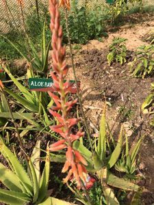
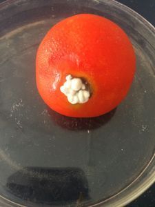
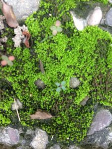
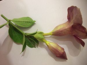
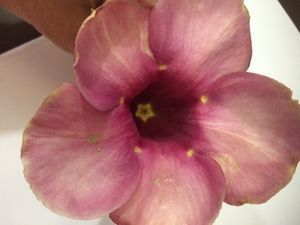
I had always imagined pollen to have thick, spiny covering called exine, which had germ pores on it. I came across Morning glory plant with pink flowers. It had bell shaped flowers, with the stamens quite deep seated. I took out the anthers, which had some staminal hairs along the filaments. I could see a larva in it. But could not see any movement in it. May be it was feeding.
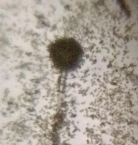
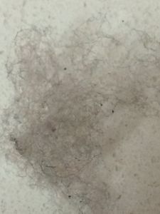
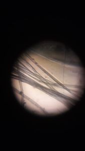
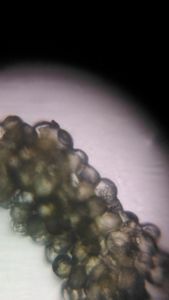
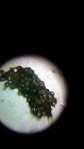
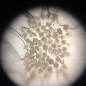
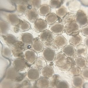
The anthers were teased to release the pollen. What I saw under a foldscope was a mass of pollen agglutinated together. Tried my best to locate some dispersed pollen, which were just a few. I observed the pollen using LED provided along with Foldscope and also without it in natural light.
The anthers were teased to release the pollen. What I saw under a foldscope was a mass of pollen agglutinated together. Tried my best to locate some dispersed pollen, which were just a few. I observed the pollen using LED provided along with Foldscope and also without it in natural light.
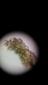
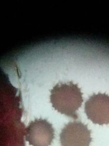
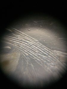
Pollen cluster in natural light
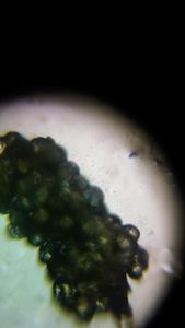
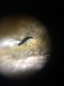
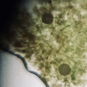
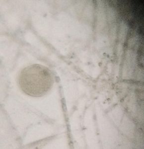
Figs. -Pollen viewed under LED and the larva or bug feeding within
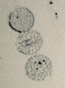
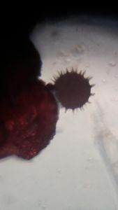
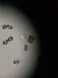
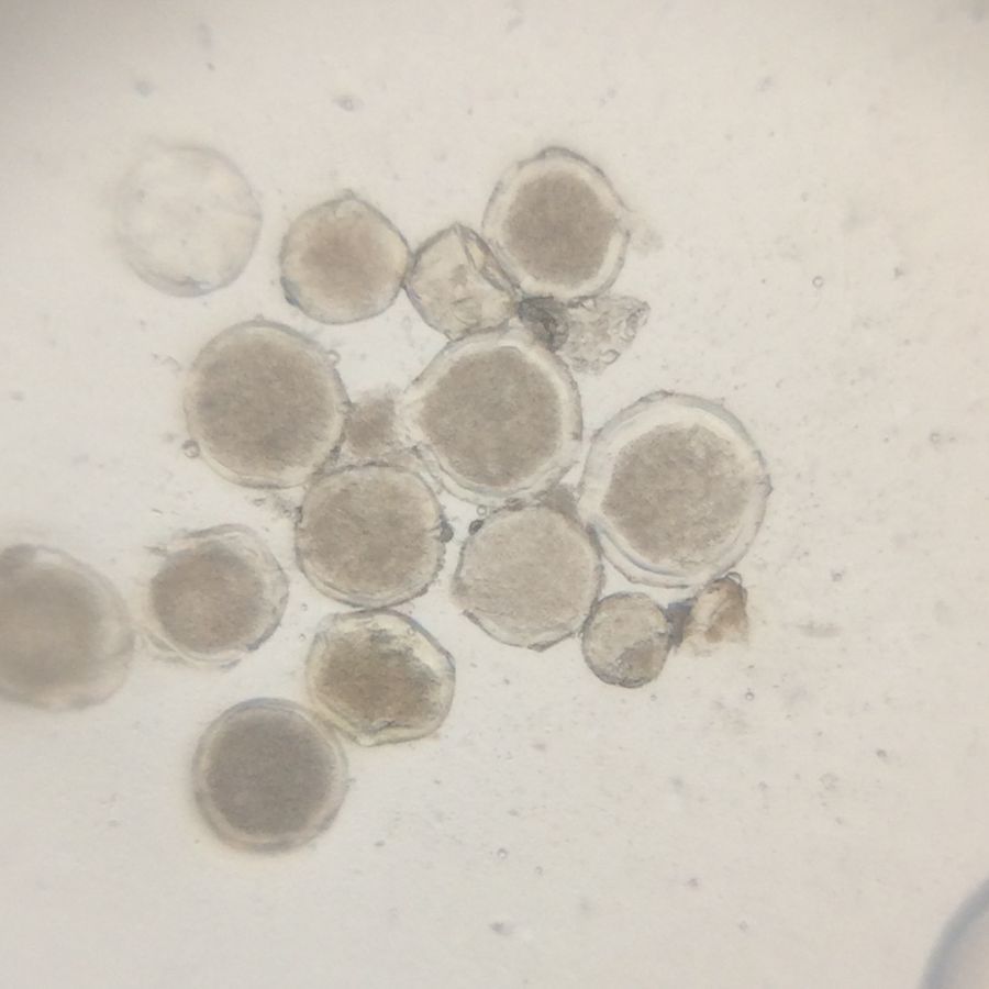
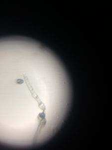
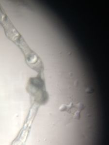
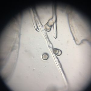
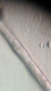
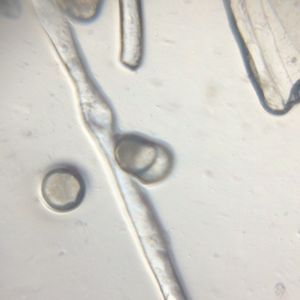
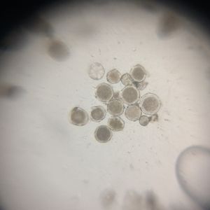
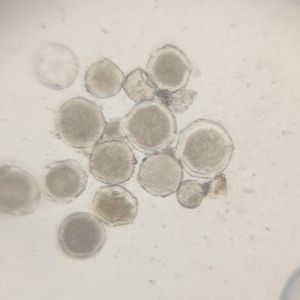
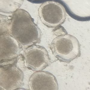
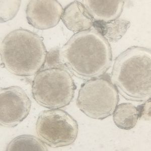
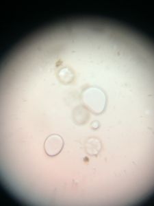
Just then an idea clicked, if I go for a wet mount? So I proceeded by taking out the pollen in a water drop. To my astonishment, the pollen separated from each other. Oh my god, these pollen are sticky. I had never thought of sticky pollen . In water these swelled up and separated from each other neatly. I could see their smooth thick coverings and even in some the germ pores, and I could easily see their pollen tubes also.
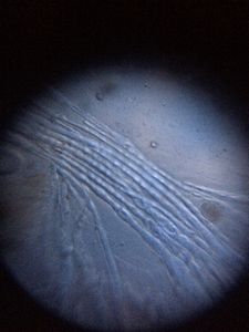
To clearly visualize the pollen tube, I used a piece of blue colored cellophane piece. With passage of time, the outer wall of pollen tended to fade out. May be it was a mucilaginous outer covering or made of a sugar, etc., which was dissolving in water, Even food reserves within the pollen grains could be spotted. And some empty pollen which had their food reserve exhausted.
This was the most amazing thing I had come to know while foldscoping.
Just then an idea clicked, if I go for a wet mount? So I proceeded by taking out the pollen in a water drop. To my astonishment, the pollen separated from each other. Oh my god, these pollen are sticky. I had never thought of sticky pollen . In water these swelled up and separated from each other neatly. I could see their smooth thick coverings and even in some the germ pores, and I could easily see their pollen tubes also.
To clearly visualize the pollen tube, I used a piece of blue colored cellophane piece. With passage of time, the outer wall of pollen tended to fade out. May be it was a mucilaginous outer covering or made of a sugar, etc., which was dissolving in water, Even food reserves within the pollen grains could be spotted. And some empty pollen which had their food reserve exhausted.
This was the most amazing thing I had come to know while foldscoping.
Sign in to commentNobody has commented yet... Share your thoughts with the author and start the discussion!
 0 Applause
 0 Comments