Comparing the peels of raw and boiled potato
 Jun 20, 2018 • 10:23 AM UTC
 India
India 140x Magnification
 Microorganisms
Ajay Prakash
Learn about the author...
21posts
0comments
3locations

View in Media Gallery
After learning foldscopy @ GGDSD, Chandigarh, these days everything at home is set to pass through FOLDSCOPE TEST . Guess what? Maa boiled potatoes for making cutlets, and the science in me asked whether cells in boiled and raw potatoes would look the same?
I picked up a potato, gently removed its peel. It was too papery (as potatoes had been cooked in microwave oven for 6 minutes —–as per my mother’s standard protocol). I soaked a part of the peel in water for about 2 hours and started my Foldscopy.
I now had the following samples for observation:
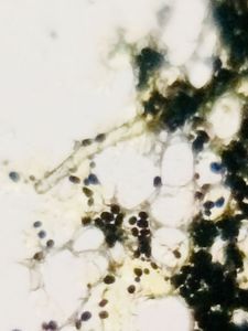
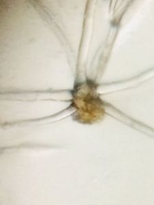
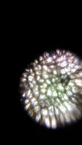
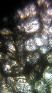
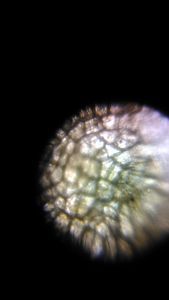
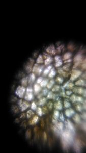
Boiled potato skin viewed from outside Boiled potato skin view from inside Boiled potato peel soaked in water for 2 hours Raw potato peel In the workshop @GGDSD, Chandigarh, potato peel had been captured by one of my fellow students, but we were not able to make out the lenticels. But now here, I could clearly mark out the dark coloured lenticels surrounded by compartively smaller cells in boiled potato skin.
I picked up a potato, gently removed its peel. It was too papery (as potatoes had been cooked in microwave oven for 6 minutes —–as per my mother’s standard protocol). I soaked a part of the peel in water for about 2 hours and started my Foldscopy.
I now had the following samples for observation:
Boiled potato skin viewed from outside Boiled potato skin view from inside Boiled potato peel soaked in water for 2 hours Raw potato peel In the workshop @GGDSD, Chandigarh, potato peel had been captured by one of my fellow students, but we were not able to make out the lenticels. But now here, I could clearly mark out the dark coloured lenticels surrounded by compartively smaller cells in boiled potato skin.
View in Media Gallery
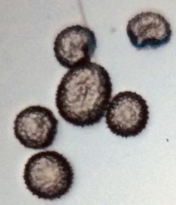
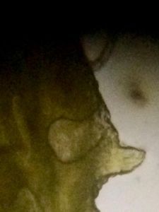
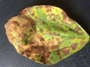
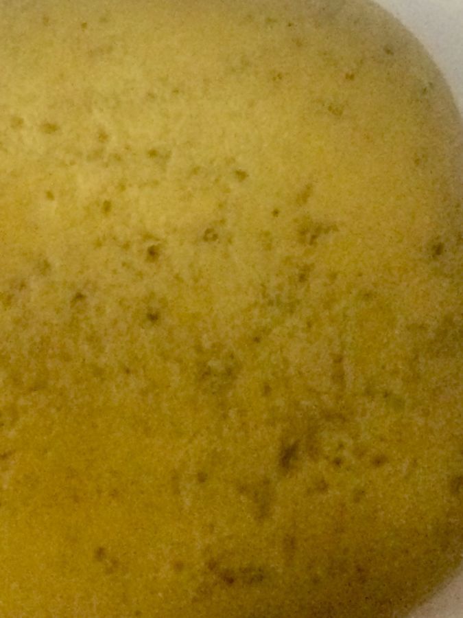
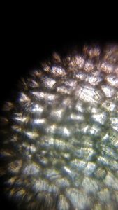
The dark colored spots on potato are the LENTICELS
Lenticels are openings consisting of cells with large intercellular spaces in the periderm of plant organs. These openings have a role in exchange of gases in potato tubers, but their discoloration leads to loss in quality and is considered a problem in post harvest technology.
Boiled potato skin viewed from outside
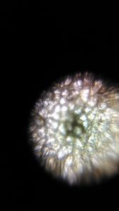

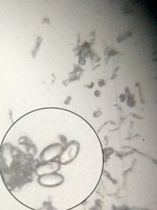
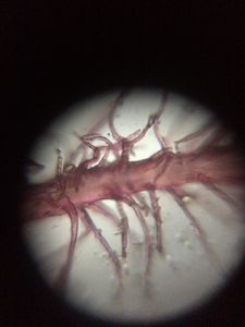
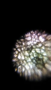
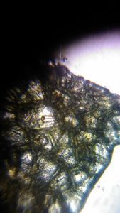
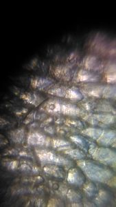
2. 2. 2. 2. Boiled potato skin view from inside
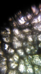
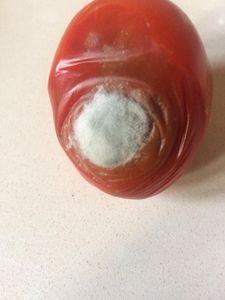
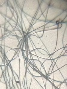
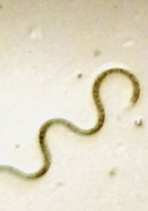
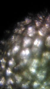
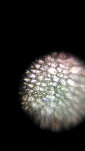
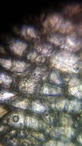
3. Boiled potato peel soaked in water for 2 hours
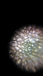
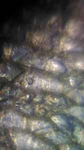
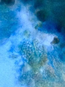
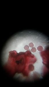
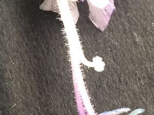
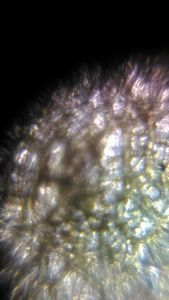
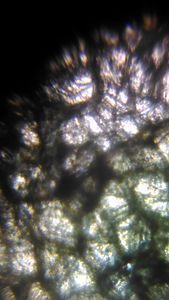
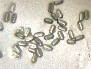
4. Raw potato peel
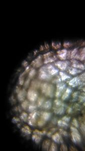
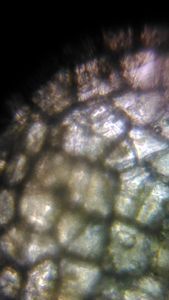
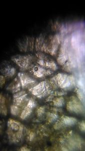
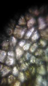
From the above pics, it can be made out that the cells in raw peel look turgid, while in the boiled peel, slightly flaccid. Also the cells surrounding the lenticels can be clearly marked out in boiled peels.
Sign in to commentNobody has commented yet... Share your thoughts with the author and start the discussion!
 0 Applause
 0 Comments