Epidermal Cyst
 Jun 21, 2018 • 3:39 AM UTC
 Unknown Location
 140x Magnification
 Microorganisms
Harshita Jaiswal
Learn about the author...
10posts
0comments
1locations

View in Media Gallery
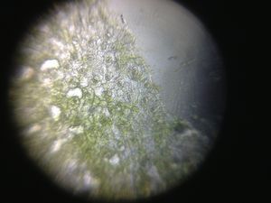
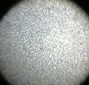
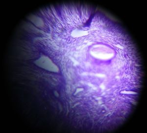
Section from an epidermal cyst shows covering epithelium and bluish flaky material inside as seen under Foldscope from Department of Pathology R D Gardi Medical College, Ujjain
Sign in to commentNobody has commented yet... Share your thoughts with the author and start the discussion!

 0 Applause
 0 Comments