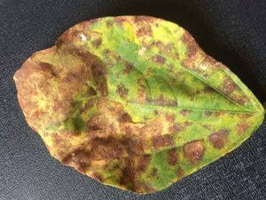
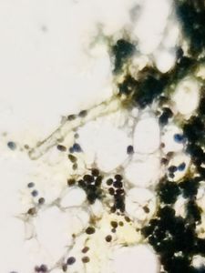
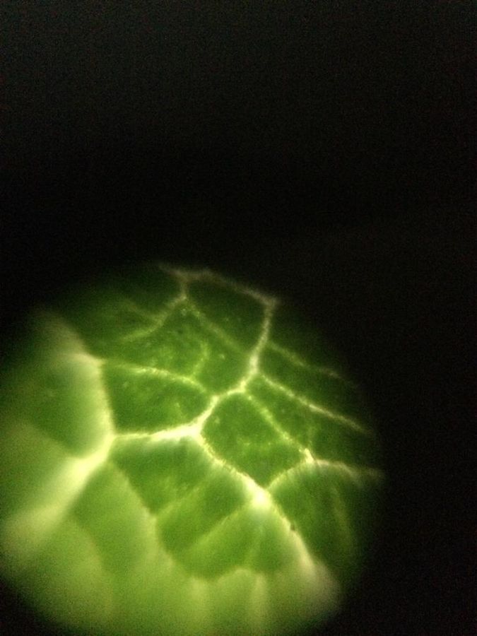
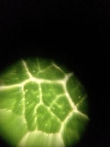
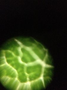
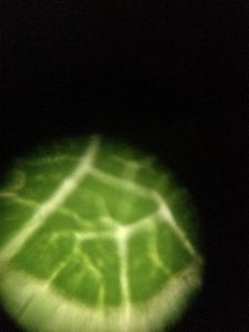
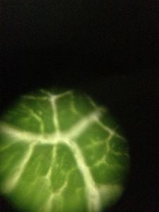
Reticulate venation in a dicot leaf
 Oct 07, 2018 • 1:04 AM UTC
 Unknown Location
 140x Magnification
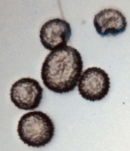
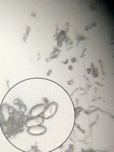
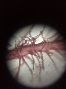
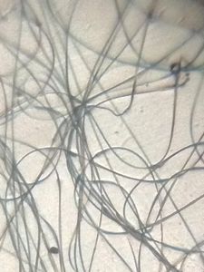
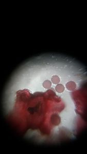
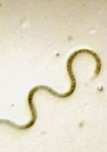
 Microorganisms
Ajay Prakash
Learn about the author...
21posts
0comments
3locations
View in Media Gallery
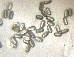
Presence of reticulate venation is an important diagnostic feature of a dicot leaf. The veins form an interconnecting network spread across the lamina. It was fascinating to observe the networking of veins under foldscope.


Sign in to commentNobody has commented yet... Share your thoughts with the author and start the discussion!
 0 Applause
 0 Comments