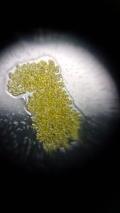
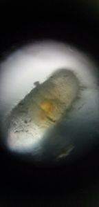
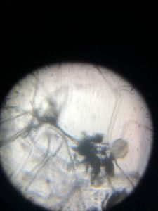
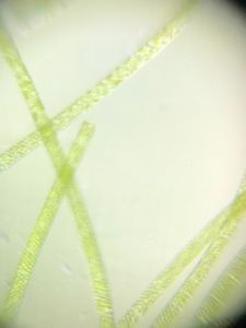
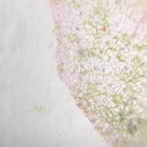
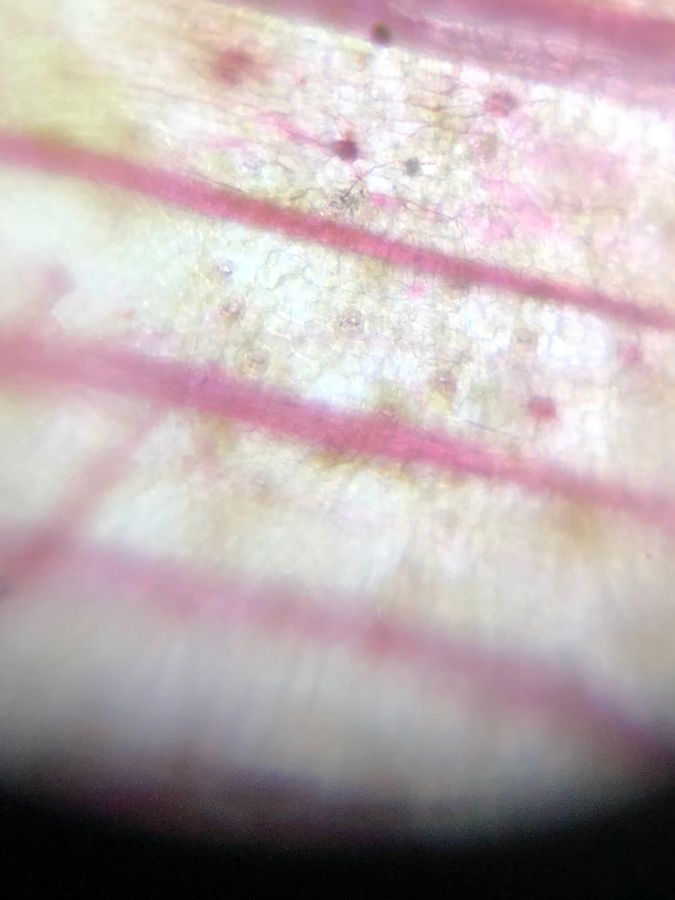
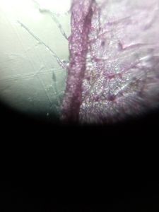
Microcosmos Logo
ExploreAll PostsAboutShop Now
ExploreAll PostsAbout
Log In or Sign Up

Orchid leaf section

Applause IconNov 21, 2018 • 10:41 PM UTC
Location IconUnknown Location
Applause Icon140x Magnification
Applause IconUnknown
User Profile
Smaranika Medhi

Junior research fellow, Bijni College, Chirang, Assam.

10posts
0comments
1locations
SINGLE IMAGE
View in Media Gallery
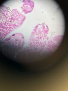
we have seen the cell nucleus, stained with saffranin.
SINGLE IMAGE
View in Media Gallery
Orchid leaf section

Sign in to commentNobody has commented yet... Share your thoughts with the author and start the discussion!

More Posts from Smaranika Medhi

Thumbnail for Xylem and phloem from plan stem
Xylem and phloem from plan stem
0 Applause0 Comments
6y
Thumbnail for Pollen observed
Pollen observed
0 Applause0 Comments
6y
Thumbnail for Ant egg observed under foldscope
Ant egg observed under foldscope
0 Applause0 Comments
6y
Thumbnail for Photoplates of national level twinning program of Bijni College,Assam and SVERI’s College of Enginnering,Pandharpur at Bijni College,Assam on 30-01-2019
Photoplates of national level twinning program of Bijni College,Assam and SVERI’s College of Enginnering,Pandharpur at Bijni College,Assam on 30-01-2019
0 Applause0 Comments
7y
Thumbnail for photoplates of national seminer on foldscope at Bijni College 0n 30th January 2019
photoplates of national seminer on foldscope at Bijni College 0n 30th January 2019
0 Applause0 Comments
7y
Thumbnail for grasshopper testis
grasshopper testis
0 Applause0 Comments
7y
Thumbnail for fungus from waste orange
fungus from waste orange
0 Applause0 Comments
7y
Thumbnail for spirogyra
spirogyra
0 Applause0 Comments
7y
Thumbnail for stomata of betel leaf
stomata of betel leaf
0 Applause0 Comments
7y
Microcosmos

About

Explore

All Posts

Brought to you by

Foldscope

© 2024 Microcosmos

Contact

Terms and Conditions

Social Media