Observation of Stained Blood Smear Using Foldscope
 Dec 11, 2018 • 12:16 AM UTC
 Unknown Location
 140x Magnification
 Microorganisms
Jitendra Satija
Learn about the author...
8posts
6comments
1locations
View in Media Gallery
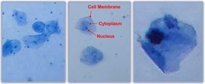
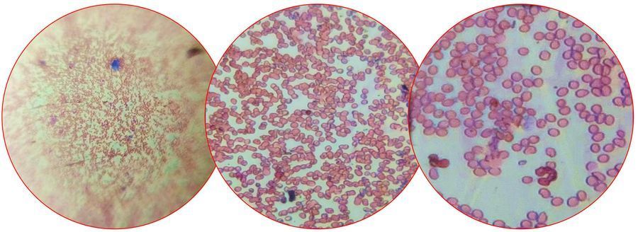
The blood smear was stained with Leishman stain and observed under the foldscope thereafter. The Leishman stain consists of two chemical groups, i.e. Azure B (basic dye) and Eosin Y (acidic dye). The erythrocytes stain pink in color due to Eosin Y as hemoglobin is eosinophilic (acidophilic), whereas the Azure B is a cationic dye which gives purple color to the nucleus and blue/light blue to the cytoplasm. This procedure and foldscopic exercise can be utilized to identify and differentiate leucocytes, malaria parasites, and Trypanosoma.
View in Media Gallery
Sign in to commentNobody has commented yet... Share your thoughts with the author and start the discussion!

 0 Applause
 0 Comments