Atharva Gautam - LGP26 - B2
 May 27, 2026 • 9:15 AM UTC
 India
India 340x Magnification
 Plants
Atharva Gautam
Learn about the author...
2posts
0comments
0locations
View in Media Gallery
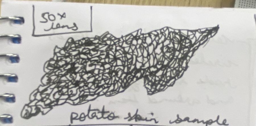
Day 3: Observing potato sample For this observation, I prepared a thin sample of potato skin and placed it carefully on the glass slide without using any stain. Since there was no staining, the structures appeared in their natural colours. At first, it was slightly difficult to focus clearly, but after adjusting the foldscope properly, I could observe interesting patterns on the potato skin.
The surface looked uneven and textured. I observed many tiny irregularly shaped cells packed closely together. Some areas appeared brighter while others looked slightly darker because of the natural thickness of the sample. I could also notice small dot-like structures scattered across the surface. The potato skin looked rough and layered under magnification. Observing an unstained sample helped me understand how the specimen naturally appears under the microscope.
We observed the sample using 50x, 140x, and 340x lenses.
Observing tomato sample For this observation, I prepared a small sample from the inner pulp of a tomato and placed it on the slide without staining. Preparing the sample was slightly challenging because the tomato pulp was soft and watery. I carefully spread a thin layer on the slide so that light could pass through properly.
When viewed under the foldscope, I could see many round and oval-shaped structures. Some appeared transparent while others looked slightly yellowish or reddish due to the natural colour of the tomato. The cells were not arranged in neat rows like onion cells; instead, they appeared more loosely spread. I also observed tiny seed-like structures and fluid-filled areas inside the sample.
The tomato sample looked very different from the potato skin because it was softer and contained more liquid. It was interesting to compare how different plant tissues appear under the foldscope even without staining.
We observed the sample at 50x, 140x, and 340x lenses.
The surface looked uneven and textured. I observed many tiny irregularly shaped cells packed closely together. Some areas appeared brighter while others looked slightly darker because of the natural thickness of the sample. I could also notice small dot-like structures scattered across the surface. The potato skin looked rough and layered under magnification. Observing an unstained sample helped me understand how the specimen naturally appears under the microscope.
We observed the sample using 50x, 140x, and 340x lenses.
Observing tomato sample For this observation, I prepared a small sample from the inner pulp of a tomato and placed it on the slide without staining. Preparing the sample was slightly challenging because the tomato pulp was soft and watery. I carefully spread a thin layer on the slide so that light could pass through properly.
When viewed under the foldscope, I could see many round and oval-shaped structures. Some appeared transparent while others looked slightly yellowish or reddish due to the natural colour of the tomato. The cells were not arranged in neat rows like onion cells; instead, they appeared more loosely spread. I also observed tiny seed-like structures and fluid-filled areas inside the sample.
The tomato sample looked very different from the potato skin because it was softer and contained more liquid. It was interesting to compare how different plant tissues appear under the foldscope even without staining.
We observed the sample at 50x, 140x, and 340x lenses.

View in Media Gallery
Sign in to commentNobody has commented yet... Share your thoughts with the author and start the discussion!
 0 Applause
 0 Comments