LGP26-B3: Day 1
 Jun 03, 2026 • 5:09 PM UTC
 India
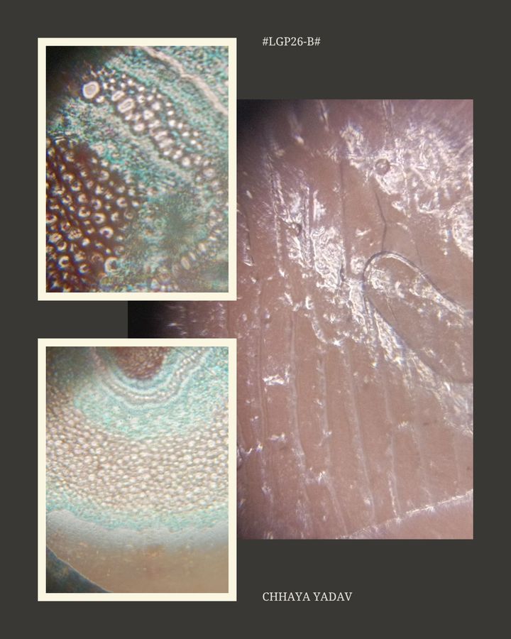
India 140x Magnification
 Plants
Chhaya Yadav
Learn about the author...
4posts
0comments
0locations
View in Media Gallery
On Day 1 of our course, we explored the fascinating world of plant cells by observing fern rhizome and onion peel samples under the foldscope. The process at first was a bit confusing but it became smooth as we moved forward. However, there were some common challenges that we saw, such as-
Using the focus ramp to focus on the sample
Sticking the sample using tape without any air bubbles
Taking the right amount of sample, thin enough for the light to pass through the sample
We observed the samples through lenses of different magnifications, i.e. 50x, 140x and 340x.
Following are the observations of each sample.
Fern Rhizome Sample:
The sample at 50x had pink, blue and purple pigment. It had block-like structure and distorted boundaries. It was overall in rough circular structure and had multi coloured smaller circles inside it.
The sample at 140x had different sized circles in darker shades of pink, purple and blue. The parts like the center and the border were more visible.
The sample at 340x had more darker shades of pigments. Some kind of hexagonal blocks were also visible.
Onion Sample:
The onion sample at 50x had a rough brick like structure. It looked transparent.
The onion sample at 140x looked like interconnected compact sticks. In this the nuclei of the cells and the cell wall were also visible.
The onion sample at 340x looked like an interconnected hexagonal structure with some bubbles moving freely.
Overall, day 1 gave us a glimpse into the hidden architecture of plant samples. I was excited to see how magnification revealed new layers of detail, turning simple samples into complex worlds.


Sign in to commentNobody has commented yet... Share your thoughts with the author and start the discussion!

 0 Applause
 0 Comments
