Day 1 : LGP26(onion)
 May 19, 2026 • 1:43 PM UTC
 India
India 50x Magnification
 Plants
Mihika Bharath
Learn about the author...
4posts
1comments
0locations
View in Media Gallery
The first time I looked at onion cells under a microscope, it felt weirdly satisfying. They looked super organized and resembled a brick wall. That's because the cells were packed tightly together.
At lower magnification (50X), I mostly saw rows of rectangular boxes, the pattern was clean and repetitive. It is kind of like graph paper made by nature.
At medium magnification (140X), the walls were much clearer and I could see the boundaries of each cell properly. The cells were transparent and were glowing because of the water inside.
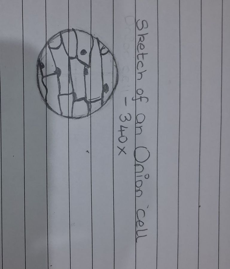
At higher magnification (340X), the details got much cooler. The cell wall was thicker and much more defined.
Getting an onion sample was harder than expected. The peel kept tearing and at first it was quite frustrating but in the end it was worth the effort.
One of the fun things that I had noticed about onion cells is that they looked much calmer compared to the fern cells. They are simple and aesthetic.
Looking at the onion sample under the microscope made me realize why they are one of the first things that people observe in biology labs. Even a tiny piece of onion holds a hidden world under the microscope. I look forward to looking at more samples
-Mihika Bharath (LGP26)
At lower magnification (50X), I mostly saw rows of rectangular boxes, the pattern was clean and repetitive. It is kind of like graph paper made by nature.
At medium magnification (140X), the walls were much clearer and I could see the boundaries of each cell properly. The cells were transparent and were glowing because of the water inside.
At higher magnification (340X), the details got much cooler. The cell wall was thicker and much more defined.
Getting an onion sample was harder than expected. The peel kept tearing and at first it was quite frustrating but in the end it was worth the effort.
One of the fun things that I had noticed about onion cells is that they looked much calmer compared to the fern cells. They are simple and aesthetic.
Looking at the onion sample under the microscope made me realize why they are one of the first things that people observe in biology labs. Even a tiny piece of onion holds a hidden world under the microscope. I look forward to looking at more samples
-Mihika Bharath (LGP26)
Sign in to commentNobody has commented yet... Share your thoughts with the author and start the discussion!
_300x300.jpeg)
 0 Applause
 0 Comments
_300x300.jpeg)