Foldscoping in the classroom: C. “Elegants” (not a typo)
 Jan 12, 2016 • 2:00 PM UTC
 Unknown Location
 140x Magnification
 Unknown
Alice Dai
Learn about the author...
12posts
6comments
1locations
View in Media Gallery
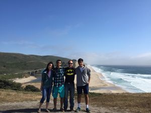
My friend Ellen Dai and I spent the last week in our research class trying to image C. Elegans on the Foldscope. We really wanted to capture the nematodes live and wriggling, and after dozens of futile attempts (mostly death by squish), we transferred a sliver of agar onto a slide and the little guys were very energetic!
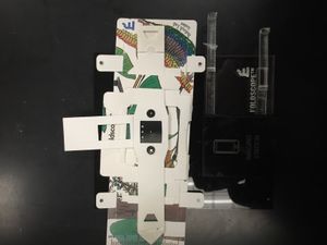
This is our setup:
This is our setup:

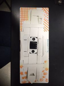
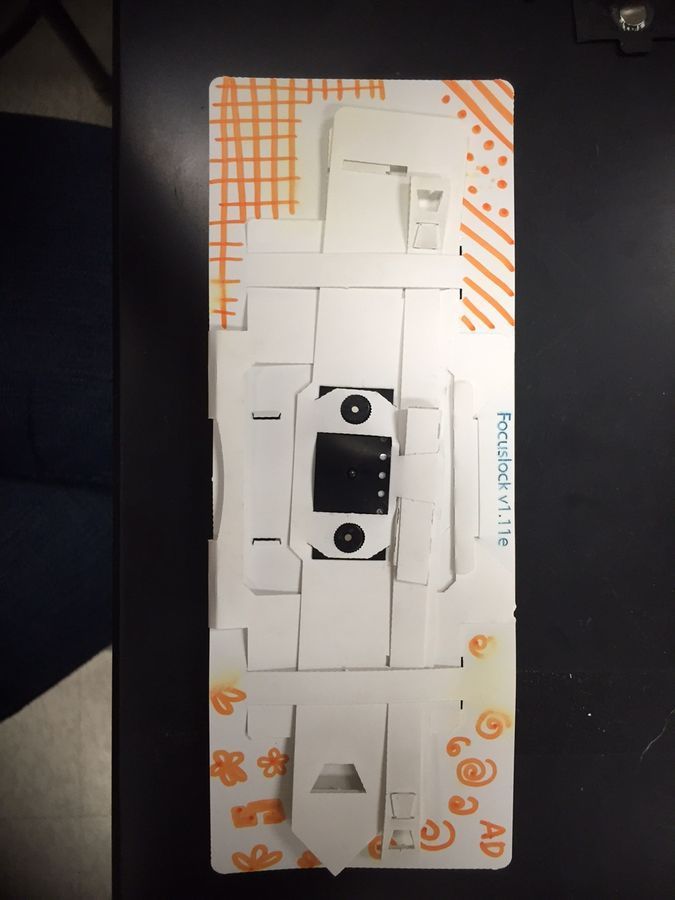
The slide on the left has C. Elegans in an agar culture (it’s clear.) I’ve decorated my Focuslock Foldscope with orange Sharpie, and the new feature on this model of the Foldscope made it a lot easier to track the C. Elegans.
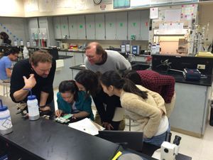
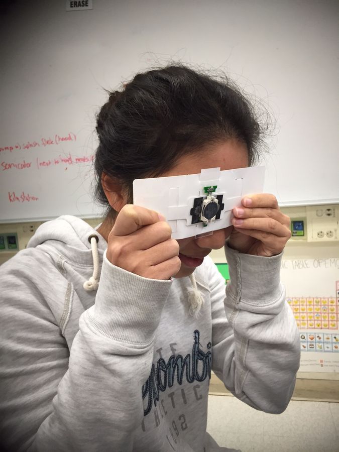
Here’s Ellen looking at the Foldscope with the dark field light module attached:
Here’s Ellen looking at the Foldscope with the dark field light module attached:
View in Media Gallery
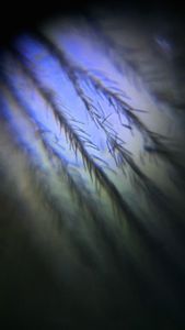
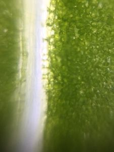
I used the dark field light module to give more contrast when imaging:
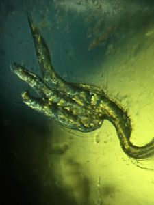
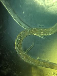
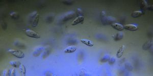
On the left is a cluster of sluggish C. Elegans (in a previous attempt to image them), and on the left is a still from a video. You can see the inner structure of the C. Elegans, which I found really fascinating. The slightly bulbous chain on the right picture look like vulva, and you can see their tapered tails as well.
And here’s a video!
And here’s a video!
Sign in to commentNobody has commented yet... Share your thoughts with the author and start the discussion!

 0 Applause
 0 Comments